-
- Figure 1
-
- Figure 2
-
- Figure 3
-
- Figure 4
-
- Figure 5
-
- Figure 6
-
- Figure 7
-
- Figure 8
-
- Figure 9
-
- Figure 10
-
- Figure 11
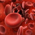
Blood
Blood is one of the body fluid that is formed of a variety of cells suspended in a fluid medium, the plasma.
Blood cells
The blood cells are grouped into three main categories: red blood cells (erythrocytes), white blood cells (leukocytes) and blood platelets (thrombocytes).
- Red blood cells (Erythrocytes)
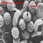
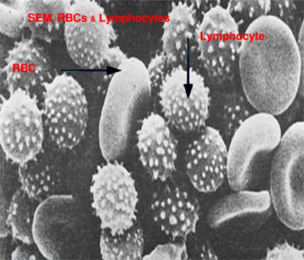
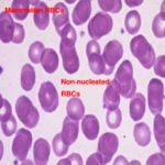
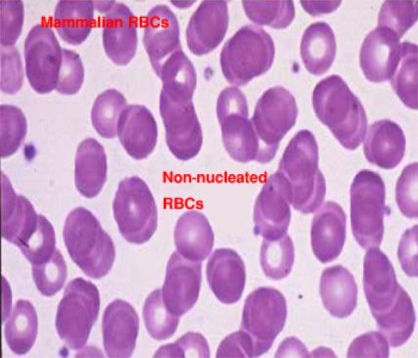
The mature red cells of domestic mammals are non-nucleated ![]() , biconcave discs.
, biconcave discs.
In pig and goat, red blood cells have no biconcavity and therefore appear as flattened discs.
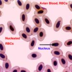
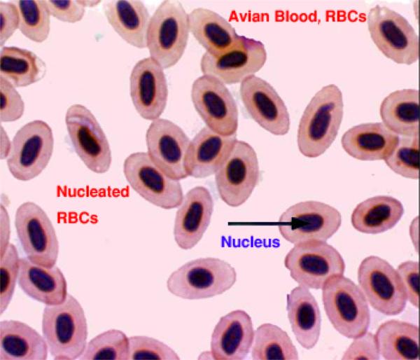
In tylopoda (camels and ilama), they are elliptical, biconcave and non-nucleated. ![]() In birds, reptiles, fishes and amphibian, they are oval, biconvex and nucleated.
In birds, reptiles, fishes and amphibian, they are oval, biconvex and nucleated.
The size of RBCs range from 4-7 mm, the largest erythrocyte is found in the dog (7mm) and the smallest in the goat (4 mm).
The number of the RBCs varies among species, in dog is about 7 million/mm3, cattle 6.3 million, goat 14 million and horse 9.5 million.
![]() With LM, the erythrocytes in a stained blood smear are stained pink due to their high content of hemoglobin. They have a central pale staining region due to their biconcave disc shape.
With LM, the erythrocytes in a stained blood smear are stained pink due to their high content of hemoglobin. They have a central pale staining region due to their biconcave disc shape.
The erythrocytes sometimes adhere to each other via their broad surface and become arranged in long chains similar to a stack of coins. This arrangement is called rouleaux.
With EM, the shape of the erythrocytes depends on the plane of section through the cell. The cytoplasmic content of erythrocytes appears electron dense due to the iron atoms of hemoglobin.
When placed in hypotonic solution (lower concentration than plasma), the RBCs swell and ruptures. This is called hemolysis.
In hypertonic solution (higher concentration than plasma), the cell volume diminishes and the cells become crenated.
The life span of erythrocytes is about 120 days. Spleen, bone marrow and liver phagocytes engulf old RBCs. The iron of the hemoglobin is reused in formation of new cells. The porphyrin portion is used to form bilirubin or bile pigment.
The erythrocytes are highly adapted to their function that is the transport of oxygen and carbon dioxide:
The plasma membrane is highly selective. It is permeable to water and electrolytes, but it is impermeable to hemoglobin.
The elasticity of the plasma membrane allows the erythrocytes to deform and pass through the smallest capillaries (2-4 mμin diameter).
The biconcave shape provides a large surface area relative to cell volume, which greatly enhance gaseous exchange.
Before release into the circulation, the nucleus is extruded and by maturity, all cytoplasmic organelles degenerate which give more space to carry more hemoglobin.
Reticulocytes
They are immature RBCs that are released into the peripheral circulation from the bone marrow. They are slightly large than mature RBCs and when stained with supravital dyes such as brilliant cresyl blue, blue-stained fine networks are seen inside their cytoplasm. This is due to ribosomal RNA still remaining in their cytoplasm. The number of reticulocytes increases in circulation after blood loss.
- II) White blood cells (leukocytes)
There are five cell types of the WBCs that are subdivided into two main groups:
- Granulocytes
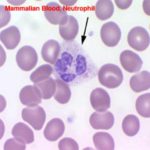
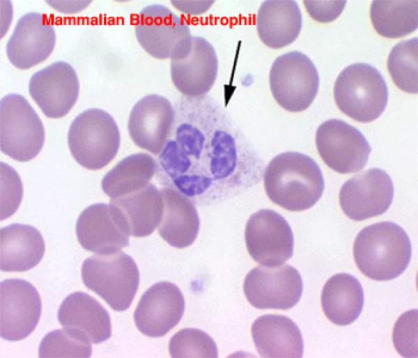
1) Neutrophils ![]()
They are the most common type of leukocytes and account for about 28 % (cattle) and 70 % (dog) of the total leukocytic count. The mature cell is about 10-12 μm in diameter.
In mature cell, the nucleus is lobulated or segmented consists of 2-5 lobes connected by fine chromatin strands. Young neutrophil has U-V- or S-shaped, non-segmented nucleus and is called band or non-segmented cells. Band cells increase in number during bacterial infection.
In female neutrophils, the quiescent X-chromosome or Barr bodies appear as a small drumstick-shaped appendage of one of the nuclear lobe. The cytoplasm contains purplish granules called azurophilic granules that are large lysosomes. Numerous smaller specific granules, are also present but they are poorly stained.
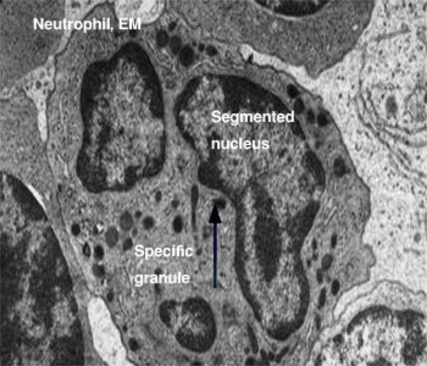
![]() With EM, neutrophil has few cytoplasmic organelles. The specific granules are relatively smaller rod-like containing bactericidal substances (phagocytins) and alkaline phosphatase. The non-specifics (azurophilic granules) are larger in size and fewer in number. They are considered lysosomes containing hydrolytic and peroxidase enzymes. Besides, glycogen granules are found. Actively migrating cells protrude pseudopodia, which are cytoplasmic extensions of the cell contain a few glycogen granules but are largely devoid of organelles.
With EM, neutrophil has few cytoplasmic organelles. The specific granules are relatively smaller rod-like containing bactericidal substances (phagocytins) and alkaline phosphatase. The non-specifics (azurophilic granules) are larger in size and fewer in number. They are considered lysosomes containing hydrolytic and peroxidase enzymes. Besides, glycogen granules are found. Actively migrating cells protrude pseudopodia, which are cytoplasmic extensions of the cell contain a few glycogen granules but are largely devoid of organelles.
Functions
Phagocytosis of invading microorganisms particularly bacteria.
They are the main WBCs type involved in acute inflammatory response. Dead leukocytes are called pus cells.
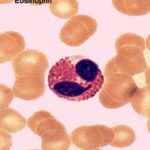
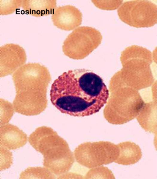
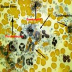
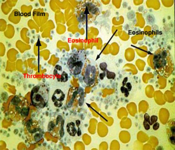
2) Eosinophils ![]()
They account for 1-6 % of the total leucocytic counts. The cell size range from 12-15 μm. Eosinophil has a bilobed, less deeply stained nucleus. The cytoplasm is packed with coarse, large, refractile, eosinophilic granules.
With EM, the cytoplasm is filled with large, ovoid, specific granules containing dense crystalloid in the long axis of the granules (dog, cat and goat). They are membrane-bound and their matrix contains a variety of hydrolytic enzymes including histaminase. Other cytoplasmic organelles such as mitochondria, rER and Golgi are relatively sparse.
Functions
The number of eosinophils in circulating blood increases during parasitic infestations and allergic conditions.
Phagocytosis of the antigen-antibody complex.
Deactivate histamine produced during inflammatory or allergic response.
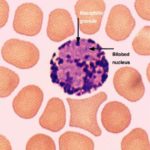
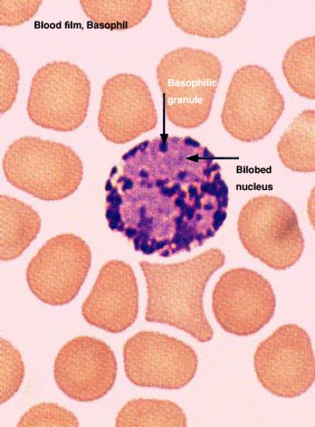
- Basophils

They are about 10-12 μm in diameter. They are the least common cell types that constitute less than 1% of the total leucocytic count. The nucleus is bilobed and completely obscured by numerous large deeply basophilic granules.
With EM, the bilobed nucleus is clearly visible and the cytoplasm is filled with membrane-bound electron dense granules.
Functions
The specific granules of the basophils contain heparin, histamine, other inactive amines, and slow reacting substance of anaphylacsis (SRS-A).
The contents of the specific granules are released by exocytosis in response to interaction of antigen with antibodies attached to the basophil cell membrane.
Heparin is anticoagulant. Histamine causes dilatation of small blood vessels and increase capillary permeability leading to exudation of fluid. SRS-A Cause contraction to the smooth muscle cells.
- Agranulocytes
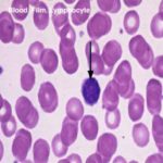
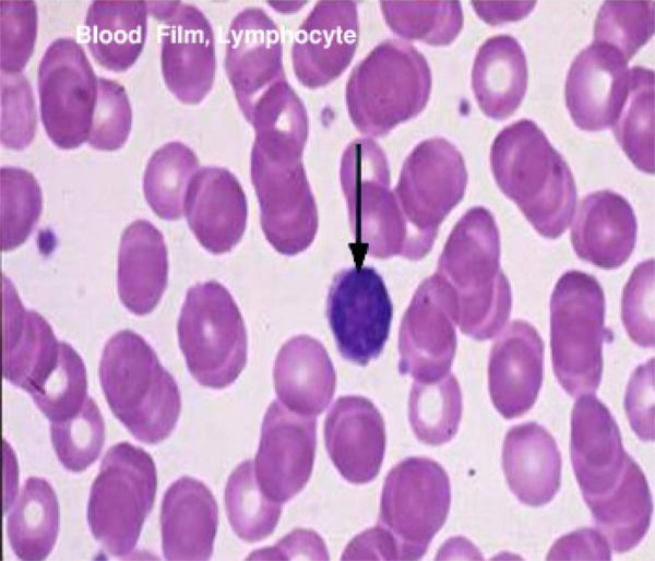
1) Lymphocytes ![]()
They are the second most common leukocytes in circulating blood. They account for 20-45% of the total leukocytic counts A round, densely stained nucleus and a relatively small amount of pale basophilic non-granular cytoplasm characterize them. According to their size, there are three types: small (6-8 μm), medium (8-10 μm) and large (10-14 μm).
With EM, the nucleus is small spherical and often slightly indented. The little cytoplasm contains a few mitochondria, a rudimentary Golgi apparatus, little or no rER and large number of free ribosomes accounting for the LM basophilia. Azurophilic granules (lysosomes) are also present.
On the basis of their functional properties, small lymphocytes are classified into two main groups: T and B-lymphocytes. Their functions will be considered with the immune system.
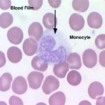
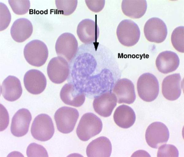
- Monocytes

They are the largest members of the white blood cell series that account for 2-10% of the total leucocytic count. They are highly motile cells and migrate into connective tissue where they are called histiocytes or tissue fixed macrophages. The large eccentricity located nucleus is bean or kidney-shaped with less densely stained chromatin than that of other leukocytes.
With LM, The cytoplasm is extensive and is filled with azurophilic granules (lysosomes. It has a frosted-glass appearance.
With EM, lysosomes are abundant, the Golgi is well developed, rER is diffuse and mitochondria are abundant than other leukocytes. It is also rich in microtubules and microfilaments. Pseudopodia are prominent reflecting their capacity for ameoboid movement and phagocytosis.
Function
The principal function of the macrophage is phagocytosis and destruction of the cellular debris.
Antigen presentation and antigen processing thus participating in both humoral and cell-mediated immune response.
III) Blood platelets (thrombocytes) ![]()
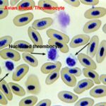
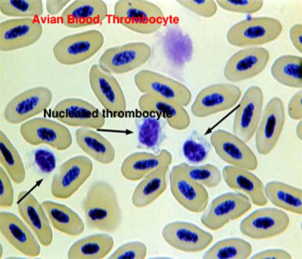
They are small, non-nucleated cells formed in the bone marrow by budding from the cytoplasm of huge cells called megakaryocytes. ![]() In birds, platelets are nucleated hence they are called thrombocytes.
In birds, platelets are nucleated hence they are called thrombocytes.
With LM, they are round or oval biconvex discs about 2-3 μm in diameter.
The cytoplasm has a purple-stained granular appearance due to its high contents of organelles. The organelles are concentrated towards the center of the cell (granulomere). The peripheral cytoplasm (hyalomere) contains microfilaments and microtubutels arranged underneath the plasmalemma. It has few organelles and is very poorly stain.
With EM, the cytoplasm is rich in membrane bound granules of two types: 1) very dense granules are sparse and contain serotonin, ADP, ATP and calcium. 2) Alpha granules which are more common and contain hydrolytic enzymes.
Functions
Blood clot formation.
Release serotonin that reduces the blood flow by constricting the damaged vessels.