-
- Figure 1
-
- Figure 2
-
- Figure 3
-
- Figure 4

-
- Figure 5
-
- Figure 6

-
- Figure 7
-
- Figure 8
-
- Figure 9
-
- Figure 10
-
- Figure 11
-

- Figure 12
-
- Figure 13
-
- Figure 14
Muscular tissue
It is one of the four basic types of tissues primarily responsible for locomotion and movement of the various body parts.
General features
- Muscle cells are long and narrow, therefore they are called muscle fibers or myofibers.
- Muscle fibers are highly specialized for contractility, which occurs due to the presence of contractile proteins within their cytoplasm.
- Muscle fibers originate primarily from the mesoderm, except the muscle of the iris, and myoepithelial cells that are ectoderm.
- Special terms are used for muscle fibers: plasmalemma = sarcolemma; cytoplasm = sarcoplasm; endoplasmic reticulum = arcoplasmic reticulum; mitochondria = sarcosomes.
- Muscle tissue is a composite tissue where it contains a minimal amount of connective tissue beside its principal cells.
- There are three types of muscle tissue: skeletal, cardiac and smooth muscles.
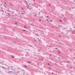
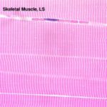
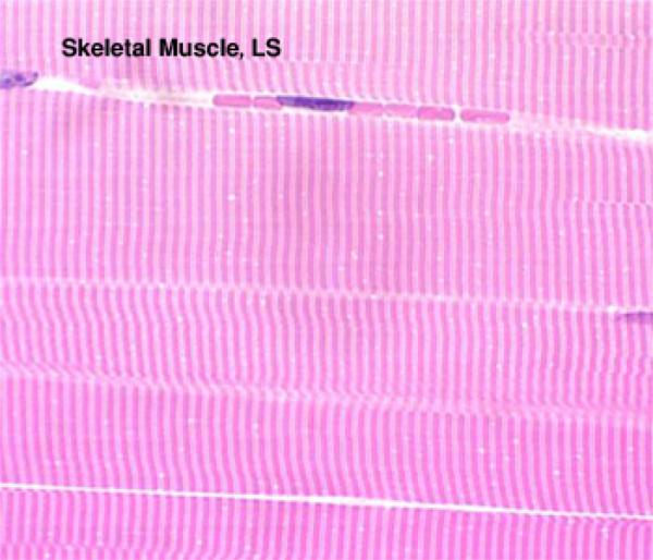
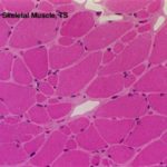
- Skeletal muscle (Striated and voluntary myofibers)
They are found in all skeletal muscles, tongue larynx, pharynx and eye. It is called skeletal because its contraction moves some parts of the skeleton; voluntary because its contraction is under conscious control, and striated because under microscope, its fiber shows alternating dark and light bands called cross-striation.
A skeletal muscle is composed of skeletal myofibers and connective tissue. A sheath of dense connective tissue called epimysium encloses the entire muscle. From the epimysium, thin collagenous septa extend inward to divide the muscle into a number of bundles or fascicles.These septa are called the perimysium. The perimysium is continuous with the endomysium that is a delicate connective tissue layer surrounds each individual myofibers.
At least five cell types are found within the bundle of the skeletal muscle: myofibers, endothelial cells, fibroblasts and myosatellite cells.
The connective tissue in between the myofibers is needed for two reasons: 1) through which blood vessels, lymphatics and nerve enter or leave the interior of the muscle. 2) At the ends of the muscle, the connective tissue elements merge to form tendons that anchor the muscle to other structures such as bone or cartilage.
Structure of skeletal myofiber
![]() At the LM level, the skeletal myofibers are extremely long, multinucleated cylindrical cells. Their diameter range from 10-100 mm and their length about 1-4 mm.
At the LM level, the skeletal myofibers are extremely long, multinucleated cylindrical cells. Their diameter range from 10-100 mm and their length about 1-4 mm.
The nuclei are oval, elongated and are located just underneath the sarcolemma.
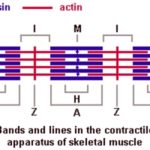
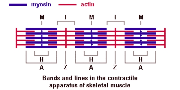
In longitudinal sections, the sarcoplasm is acidophilic and crosses striated with alternating dark and light-staining transverse bands.
Under the polarized light, the dark-staining bands are called anisotropic or A-bands because polarized light is reflected unequally when passed through it. The light-staining bands are called isotropic or I-band because polarized light is reflected equally when passed through it. Dark lines termed Z-lines or Z-disc bisects the I-bands. The center of each A-band contains a paler region called H-zones or H-band.
The distance between two successive Z lines is called sarcomere that is the contractile unit of the skeletal muscles.
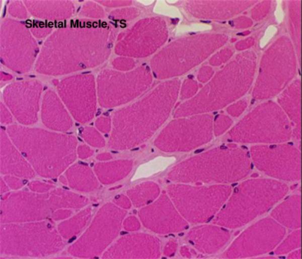
![]() In cross sections, the skeletal myofibers appear oval, spherical or polygonal with their nuclei are peripherally located.
In cross sections, the skeletal myofibers appear oval, spherical or polygonal with their nuclei are peripherally located.
![]() At the EM level, the skeletal myofiber is surrounded by sarcolemma with the usual trilaminar appearance. The sarcoplasm is filled with myofibrils arranged parallel to the long axis of the cell. Each myofibril has cross-striations and they are arranged with their cross striations in register so as to give the LM appearance of regular cross-striations along the muscle fiber. The myofibrils are separated by a small amount of sarcoplasm containing rows of mitochondria.
At the EM level, the skeletal myofiber is surrounded by sarcolemma with the usual trilaminar appearance. The sarcoplasm is filled with myofibrils arranged parallel to the long axis of the cell. Each myofibril has cross-striations and they are arranged with their cross striations in register so as to give the LM appearance of regular cross-striations along the muscle fiber. The myofibrils are separated by a small amount of sarcoplasm containing rows of mitochondria.
![]() The myofibrils are found to be composed of smaller units called the myofilaments that are of two types: myosin (thick) and actin (thin) filaments.
The myofibrils are found to be composed of smaller units called the myofilaments that are of two types: myosin (thick) and actin (thin) filaments.
The thick filaments are composed mainly of protein myosin and are arranged parallel to each other in the A-band. They are maintained in parallel by their attachment to a disc-like zone called the M-band that is located in the center of H-band.
The thin filaments are composed mainly of protein actin that is associated with two other proteins, tropomyosin and troponin. They are attached to both sides of the Z-lines to form the I-band.
The A-bands are electron-dense and appear dark because they contain two types of filaments.
The H-bands and I are of low electron density and appear light because the thin and thick filaments do not overlap one another.
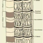
The sarcolemma gives rise to tubular extensions (T-tubules) that extend transversely in the sarcoplasm to surround each myofibril at the region of A-I junctions.
Each tubule is associated with two dilated cisternae of the sarcoplasmic reticulum to form a structure called triad. The T-tubules transmit nerve impulses from the outer sarcolemma to the sER cisternae leading to release of Ca++ ions that are necessary for myofibers contraction.
Other cytoplasmic organelles include small inactive Golgi, dense glycogen and well-developed sER.
Red and white fibers
- Red myofibers
They have a red color in fresh state due to their high myoglobin contents that is an oxygen-storage molecules similar to hemoglobin. They are smaller in diameter and contain abundant cytochromes and mitochondria. They have a rich blood supply. They are aerobic and predominate in muscle that can sustain their contractile activity for a long time.
- White myofibers
They are larger in cross-section. They contain few mitochondria and cytochromes and relatively little myoglobin hence, they are white in fresh state. They have a relatively poor blood supply. The are rich in glycogen. They are anaerobic white fibers and constitute muscles that perform intense but sporadic contraction. The white fibers contract more rapidly but they also fatigue more rapidly.
In most mammalian species, the skeletal muscles are composed of a mixture of red and white fibers. In domestic poultry, some muscles are red such as leg muscles, and others are white such as pectoral muscle. In wild fowl, the pectoral muscles are composed of red fibers that are capable of sustained flight.
- Cardiac muscle (Striated and involuntary)
It is called cardiac because it constitutes most of the heart although some cardiac muscle can also be found in the wall of pulmonary vein and vena cava.
Cardiac myofibers are striated, however, the striation is less distinct than that of the skeletal myofibers due to: irregular branching shape of the fiber, the less myofibrils content and the abundance of non-contractile sarcoplasm. They are involuntary, they contract spontaneously without any nerve supply. The rate this inherent rhythm can be modulated by autonomic and hormonal stimuli.
Structure of cardiac muscle
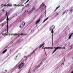
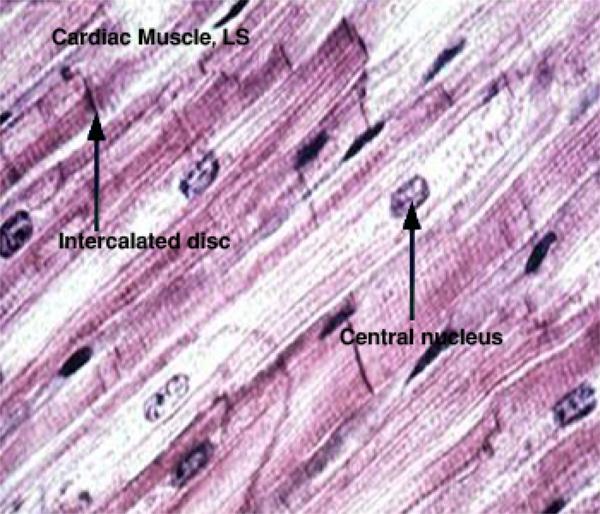
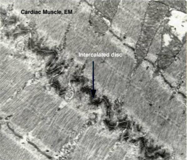
![]() At the LM level, the cardiac myofibers are long (50-100 mm), cylindrical cells that are branched and anastomosed forming a network. They are traversed at intervals by dark-staining structures called intercalated discs that extend across the fiber in a stepwise manner.
At the LM level, the cardiac myofibers are long (50-100 mm), cylindrical cells that are branched and anastomosed forming a network. They are traversed at intervals by dark-staining structures called intercalated discs that extend across the fiber in a stepwise manner.
Most of the cells have one nucleus and at most two nuclei. The nuclei are oval and centrally located within the cell. Like the skeletal muscles, the cytoplasm is acidophilic and striated consisting of an alternated dark and light bands.
The cells are surrounded by delicate connective tissue containing fibroblasts, pericytes and dense capillary network necessary to meet their high metabolic demands. Myosatellite cells are absent.
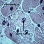
![]() In TS, the cardiac muscle fibers are spherical or oval with aregular diameter of about 20 mm containing single central nuclei.
In TS, the cardiac muscle fibers are spherical or oval with aregular diameter of about 20 mm containing single central nuclei.
At the EM level, the fine structure of the cardiac muscle is similar to that of the skeletal muscle except:
- Cardiac myocytes has a less extensive sarcoplasmic reticulum and does not form dilated terminal cisternae.
- The T tubules are wider than those of the skeletal myofibers and penetrate the cardiac myocytes at the level of Z-lines and not at the A-I junctions.
- Each tubule is associated with single cisternae of sarcoplasmic reticulum forming a structure called diad.
- Mitochondria are numerous with closely packed cristae rich in oxidative enzymes.
- The sarcoplasm contains larger amount of glycogen.
- The perinuclear sarcoplasm of cardiac myofibers contain a large number of secretory granules that have recently been proven to be the source of polypeptide hormone called ANP (Atrial Natriuretic Peptide).
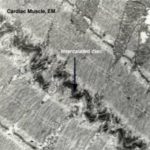
- The cardiac myofibers are formed of several cells connected end-to-end by intercalated discs
 . Each intercalated disc crosses a cardiac fiber at the Z-line level in a stepwise manner. It consists of three types of cell junctions: fascia adherens, desmosomes and gap junction. The ntercalated discs bind the cell, transmit forces of contraction and provide areas of low electrical resistance for the spread of excitation throughout the myocardium.
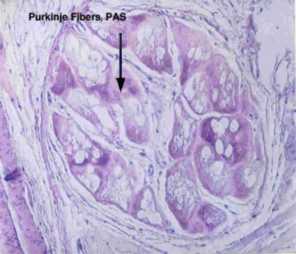
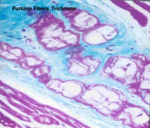
Purkinje fibers ![]()
![]()
They are modified cardiac muscle fibers designed for rapid conduction of nerve impulses. They differ from the ordinary cardiac muscle fibers in the following aspects:
- They are larger in size.
- The nucleus is smaller and eccentricity situated.
- The cytoplasm is paler, vacuolated because it is rich in glycogen.
- The myofibrils are fewer and concentrated at the periphery.
- The T tubules are absent.
3) Smooth muscle (Visceral muscle)
Smooth muscles are found in the walls of hollow viscera and blood vessels. It is called smooth because it has no cross striations, involuntary because its contraction can not be elicited at will and visceral because they are found in visceral organs.
Structure of smooth myofibers
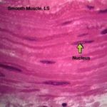
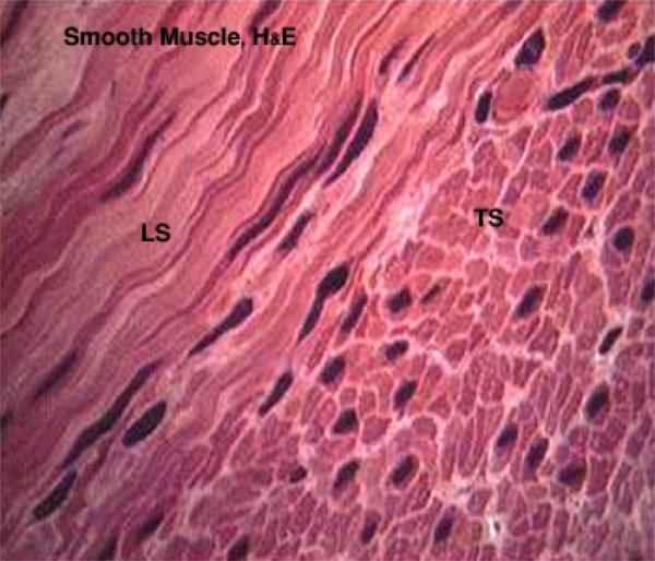
![]() At the LM level, the smooth muscle cell is elongated, spindle-shaped with pointed ends. It has a diameter of 3-10 mm with length ranges between 30-500 mm.
At the LM level, the smooth muscle cell is elongated, spindle-shaped with pointed ends. It has a diameter of 3-10 mm with length ranges between 30-500 mm.
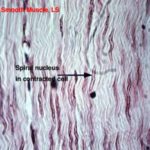
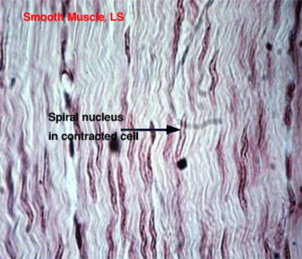
The cytoplasm is acidophilic and contains only one nucleus. The nucleus is elongated and centrally located in the cytoplasm at the widest part of the cell. During contraction, the nuclei may appear spiral in shape ![]() .
.
The smooth muscle fibers are bound together into irregular branching bundles. Within the bundles, individual muscle fibers are arranged parallel to each other with
the thick part of the cell lying against the thin parts of adjacent cells. A sheath of delicate connective tissue containing capillaries and few nerve fibers invests each muscle bundle.
![]() In cross sections, the spindle shaped cells is sectioned at different levels along their length. The cells appear spherical or oval with a differing diameter. Nuclei are only included when the fiber cut through their widest portion and is located centrally.
In cross sections, the spindle shaped cells is sectioned at different levels along their length. The cells appear spherical or oval with a differing diameter. Nuclei are only included when the fiber cut through their widest portion and is located centrally.
At the EM level, the plasma membrane contains numerous flask-shaped invaginations called caveole. They are regarded as the probable counterparts of T tubules. Mitochondria, Golgi saccules, glycogen and small amount of rER are largely confined to the perinuclear cytoplasm.The sER is poorly developed consisting of narrow tubules with no terminal cistenae. There are no T tubules.
The cytoplasm is filled with parallel thin (actin) and thick (myosin) filaments. The filaments do not have the arrangement seen in the sarcomeres.
Intermediate filaments (desmin, vimentin and synemin) are also located within the cytoplasm where they are attached to electron-dense structures known as dense bodies that represent the counterpart of Z lines.
Nexus-like junctions that facilitate the spread of excitation throughout visceral muscle attach the cells. Desmosomes are also found where they bind adjacent cells and provide anchorage points for actin and myosin.
Functions
- Contraction of the smooth muscle is an inherent property however, it can be modulated be autonomic nervous system.
- The smooth muscle maintains prolonged partial contraction (tonus) in the wall of arterioles that is necessary to keep normal blood pressure in the blood capillaries.
- In the small intestine, the smooth muscle cell undergoes continuous rhythmic constrictions passing along the tract propelling the lumenal contents distally.
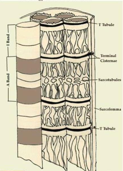
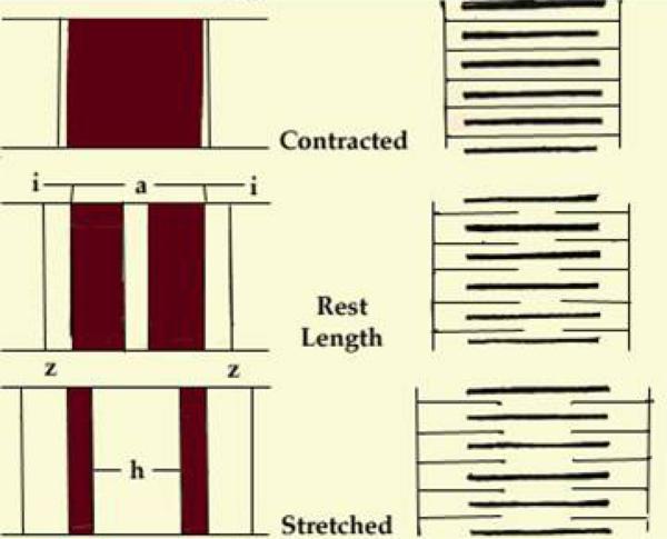
Contraction of skeletal muscle (Sliding filament theory) ![]()
![]()
In relaxed sarcomer, the thick (myosin) filaments are located in the center region forming the A-band. Both ends of the thick filaments are free.
The thin filaments (actin) have only one end free and the other end is attached to a Z line. Their free ends extend toward the middle of the sarcomer to interdigitate with the thick filaments.
When the muscle fiber is stimulated to contract sliding of the thin and thick filaments occurs without change in the length of either filament.
The Z lines of all sarcomeres are drawn toward each other resulting in contraction.