-
- Figure 1
-
- Figure 2

-
- Figure 3
-
- Figure 4
-
- Figure 5
-
- Figure 6
-
- Figure 7
-
- Figure 8
-
- Figure 9
-
- Figure 10
-
- Figure 11
-
- Figure 12
-
- Figure 13

-
- Figure 14
-
- Figure 15
-
- Figure 16
-
- Figure 18
-
- Figure 18
-
- Figure 19
-
- Figure 20
Nervous System
The nervous system is divided into two main branches: the peripheral nervous system (PNS) and central nervous system (CNS).
The central nervous system comprises brain (cerebrum, cerebellum and brain stem), and spinal cord.
The peripheral nervous system comprises cranial and spinal nerves, nerve endings and ganglia.
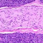
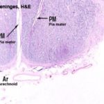
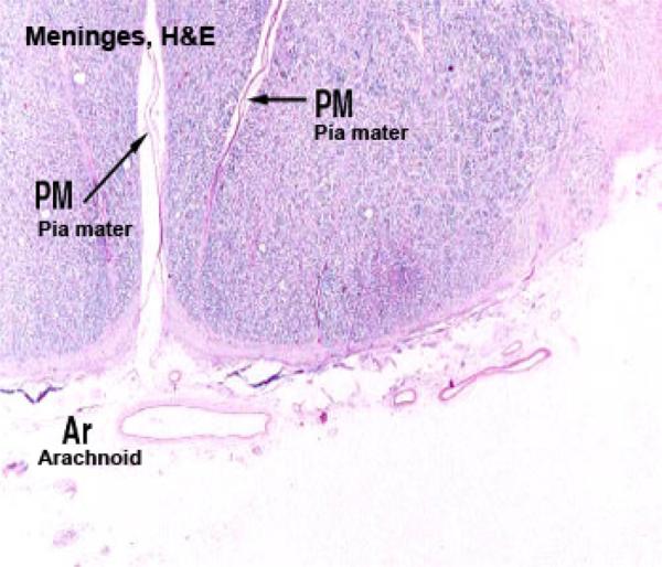
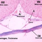
The brain and spinal cord are invested by three layers of connective tissues called the meninges that provide protection against traumatic forces. These layers are dura mater, pia mater and arachnoid.
Dura Mater
The dura mater is a dense fibroelastic connective tissue layer.
In the cranium, the dura is fused with the periosteum.
Around the spinal cord, the dura mater is suspended from the periostium of the spinal canal by denticulate ligaments.
The space between the dura and the overlying periostium is called epidural space and is filled with loose connective tissue, fat and a venous plexus.
Arachnoid
The arachnoid consists of a delicate connective tissue layer of fine collagenous fibers in contact with the dura mater, and arachnoid trabeculae which are spider web-like thin strands of membranes lined by flattened fibroblasts.
The arachnoid trabeculae traverse the subarachnoid space that is located between the connective tissue layer and the pia mater.
The subarachnoid space is filled with cerebrospinal fluid. As vessels penetrate the CNS, an extension of the subarachnoid space extends around the vessels forming perivascular space.
Pia Mater
It is a delicate vascular connective tissue layer containing fibroblasts, collagenous fibers and the processes of underlying astrocytes resting on the basal lamina that covers the nervous tissue.
Central Nervous System
The C.N.S comprises the brain (cerebrum and cerebellum) and the spinal cord.
Within the CNS, internal and external stimuli are analyzed and integrated to produce appropriate responses in various effector organs.
On gross examination, the CNS is made up of gray mater and white mater.
The gray matter consists mainly of neuron cell bodies while the white matter consists of bundles of myelinated and non-myelinated axons.
It is the lipid in the myelin sheaths of the axons that gives the white appearance of the white matter.
Brain
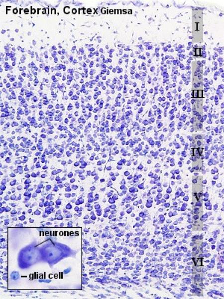
The brain is subdivided into cerebrum, cerebellum and brain stem.
The cerebrum is divided into two cerebral hemispheres by deep longitudinal fissure.
The outer surface of the cerebrum is convoluted and has many gyri and sulci.
The cerebral hemisphere consists of convoluted outer cortex of gray matter overlying inner medulla of white matter.
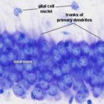
Types of neurons in the cerebral cortex:
Pyramidal cells
The pyramidal cells are pyramidal in shape with their sizes varies greatly.
The smallest pyramidal cells are located superficially.
However, the large cells (Betz cells) are located deeper within the cortex.
The cell apex is directed outward and it has a thick branching dendrite towards the surface.
In addition, short dendrites arise from the edges of the base and extend laterally. A thin axon arises from the base of the cell and passes into the underlying white matter.
- Stellate (granule) cells
They are small neurons with short vertical axon and several short branching dendrites giving the cell body a star shape.
- Cells of Martinotti
They are small polygonal cells superficially located within the cortex.
They have short dendrites with a single axon extends toward the surface and divides to run horizontally.
- Fusiform cells
They are spindle-shaped cells arranged at right angles to the surface.
The axon arises from the side of the cell body and extends superficially. Dendrites arise from each cell end and pass vertically into either deeper or superficial layers.
- Horizontal cells of Cajal
They are small spindle-shaped cells oriented parallel to the surface. They are superficially located, their axons extend laterally to synapse with the dendrites of pyramidal cells.
Layers of Cerebral Cortex
The cerebral cortex is made up of six layers that merge with one another without any clear line of demarcation.
Molecular or Plexiform Layer: It is the outer most layer and is formed of dendrites and axons of the underlying neurons. It also contains few neuroglia cells and horizontal cells of Cajal.
Outer Granular Layer: consists of dense population of small pyramidal and stellate cells as well as axons and dendrites from deeper layers.
Outer Pyramidal Layer consists of medium-size pyramidal cells.
Inner Granular Layer: consists of densely packed stellate cells.
Inner Pyramidal (ganglionic) Layer: consists of large pyramidal cells and smaller number of stellate cells.
Multiform or Fusiform Layer contains fusiform cells in its deeper layer. Other cell types such as small pyramidal cells, cells of Martinotti as well as stellate cells are located superficially.
The white matter of the cerebrum is made up of ascending and descending tracts of sensory and motor fibers respectively.
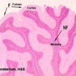
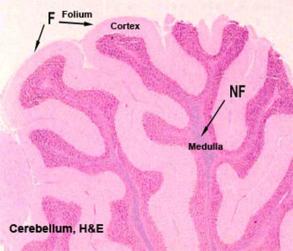
The surface of the cerebellar cortex forms convoluted folds called folia.
Each folium has an outer cortex of gray matter and an inner medulla of white matter. The cerebellar cortex consists of three layers:
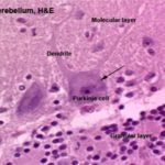
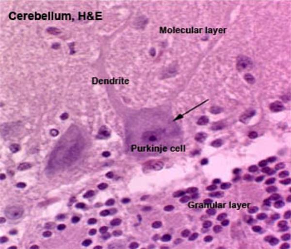
Outer Molecular layer: is located most superficially and consists of numerous dendrites with few small stellate and basket-like neurons.
Intermediate Purkinje (Piriform) Layer: ![]() It consists of a single layer of large, flask-shaped neurons called Purkinje cells.
It consists of a single layer of large, flask-shaped neurons called Purkinje cells.
Each Purkinje cell has a huge cell body with a single axon extends down and extensively branching dendrites extend into the outer molecular layer.
Inner granular Layer: situated adjacent to white matter and consists of densely packed small neurons with heterochromatic nuclei.
Functions of Cerebellum
Co-ordinate muscular activity.
Maintain posture and equilibrium.
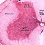
In cross section, the gray matter is located centrally taking a butterfly or H-shape.
A central canal containing cerebrospinal fluid and lined by ependymal cells lies in the center of gray matter.
The H-shaped gray matter has two dorsal horns; two ventral horns and two lateral horns.
The ventral horns are more prominent ![]() and contain the cell bodies of large motor neurons .
and contain the cell bodies of large motor neurons .
The dorsal horns are much less prominent and contain the cell bodies of small sensory neurons.
The lateral horns in the thoracic and lumber regions contain the cell bodies of preganglionic sympathetic efferent neurons. The cervical and lumber regions of the spinal cord have much greater diameter with a much more extensive gray matter volume.
Theses regions give the extensive sensory and motor innervation of the fore and hind limbs.
The white matter of the spinal cord is located peripherally around the gray matter.
It consists of ascending (sensory) and descending (motor) tracts.
The volume of the white matter increases from the sacral to cervical region.
Externally, the white matter is divided bilaterally by deep ventral median fissure and dorsal median septum.
On each side, a dorso-lateral sulcus marks the line of entery of the dorsal nerve roots.
The site of exit of the ventral nerve roots is also marked by two ventrolateral sulci.
The triangular area of the white matter between the dorsal horns represents the ascending dorsal columns that convey fibers for the senses of vibration, proprioception and touch to the brain.
The ventrolateral white matter is made up of various ascending (sensory) and descending (motor) tracts such as lateral spinothalamic tract (pain and temperature), ventral spinothalamic tract (light touch).
Peripheral Nervous System
The PNS comprises cranial and spinal nerves, nerve endings and ganglia.
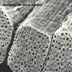
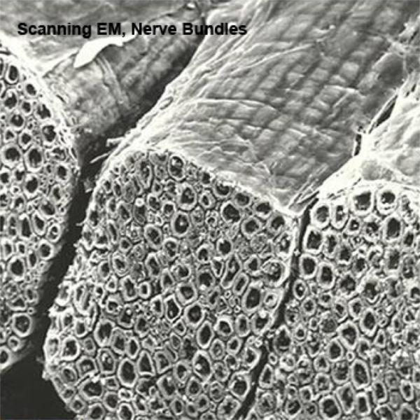
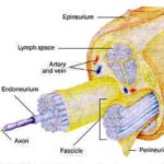
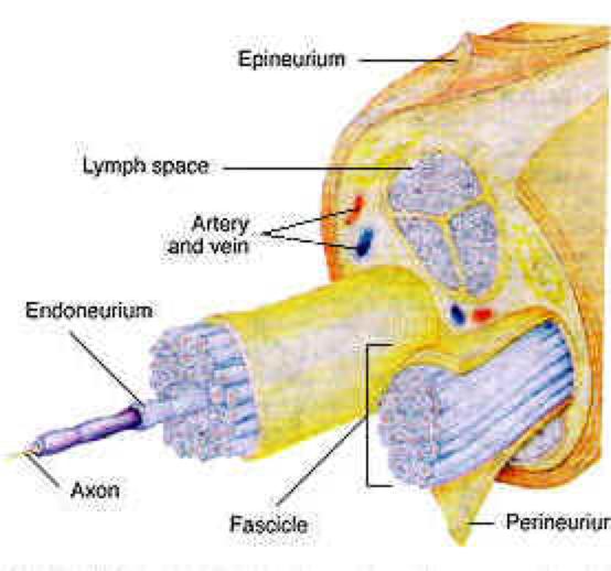
Peripheral Nerve Fibers
The term nerve fiber refers to either a single myelinated axon with its suroounding Schwan cells or to a single non-myelinated axon.
The term nerve ![]()
![]() refers to a collection of axons (thousands) each invested by neurolemmocytes and all are surrounded by connective tissue.
refers to a collection of axons (thousands) each invested by neurolemmocytes and all are surrounded by connective tissue.
Types of nerve fibers
Afferent (sensory) Nerve Fibers
The afferent nerve fibers transmit nerve impulses from receptors in the body tissues and organs to CNS.
Afferent somatic nerve fibers are distributed to skin, skeletal muscle and bone.
Afferent visceral nerve fibers are distributed to visceral organs and glands.
Efferent (motor) fibers
The efferent nerve fibers transmit nerve impulses from the CNS to various tissues and organs.
Efferent somatic (voluntary) nerve fibers innervate skin, skeletal muscles and joints.
Efferent visceral (involuntary) nerve fibers innervate cardiac and smooth muscle and glands.
The efferent visceral fibers form the autonomic nervous system (ANS).
The autonomic pathway consists of two neurons, the first neuron is located in the CNS (brain and spinal cord) and is called preganglionic neuron. The cell body of the second neuron is located in autonomic ganglia.
The ANS has two main divisions: sympathetic branch and parasympathetic branch.
In parasympathetic branch, the cell bodies of the preganglionic neurons are located in the brain and sacral region of the spinal cord (cranio-sacral).
In sympathetic branch, the cell bodies of the preganglionic neurons are located in the thoracic and lumber regions of the spinal cord (thoraco-lumber).
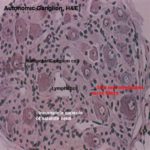
Peripheral Ganglia
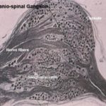
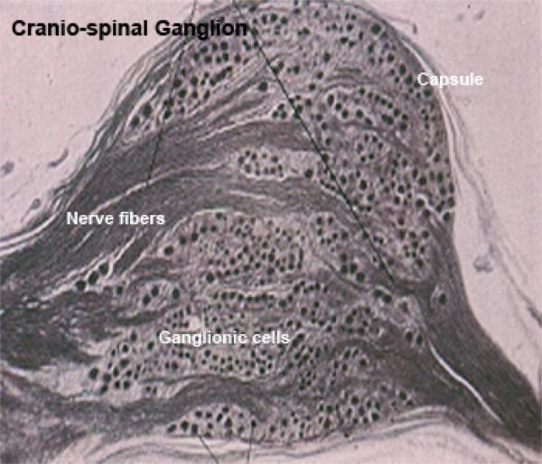
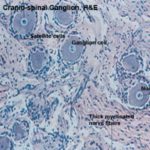
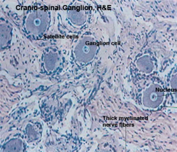
Ganglia are discrete aggregations of neuron cell bodies located outside the CNS. There are two types of ganglia: craniospinal and autonomic ganglia.
They are found along the course of some cranial nerves and all spinal nerves.
They are fisiform in shape and are surrounded by a connective tissue capsule of collagenous and reticular fibers.
The neurons are pseudounipolar.
The cell bodies are spherical with different sizes ranging from 15-120 mm.
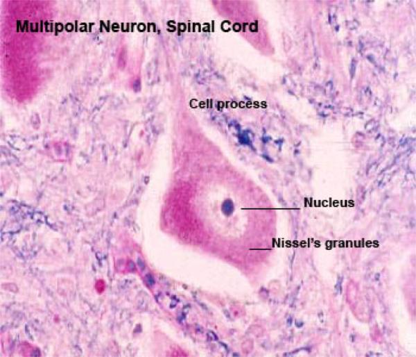
The nuclei are spherical large euchromatic with prominent nucleoli and are centrally located.
The cytoplasm contains basophilic (Nissle’s) granules. The cells are arranged in clusters that are located peripherally.
Each cell body is surrounded by a complete layer of flattened satellite cells that provide structural and metabolic support. Blood vessels, dendrites and bundles of axons are found between the ganglionic cells.
The nerve fibers are thick and myelinated.
They are similar to craniospinal ganglia being made up of a connective tissue framwork containing ganglion and capsule cells. However, they differ in:
They have a thin connective tissue capsule.
The ganglion cells are multipolar and appear polygonal or irregular in shape.
The cell bodies are smaller and have the same size (30 mm).
The ganglion cells are not arranged in clusters and are randomly scattered throughout the ganglia.
The nuclei are eccentric in position.
The ganglion cells are surrounded by incomplete capsule of satellite cells.
The nerve fibers are thin and non-myelinated.
The sympathetic ganglia are interconnected to form bilateral sympathetic chains (trunk) located along the ventral surface of the vertebral column.
The parasympathetic ganglia are located within or near the effector organs.
The cell bodies may be aggregated as discrete ganglia but more common, parasympathetic ganglia appear as few cell bodies aggregated together to form minute ganglia scattered in the supportive connective tissue.
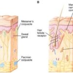
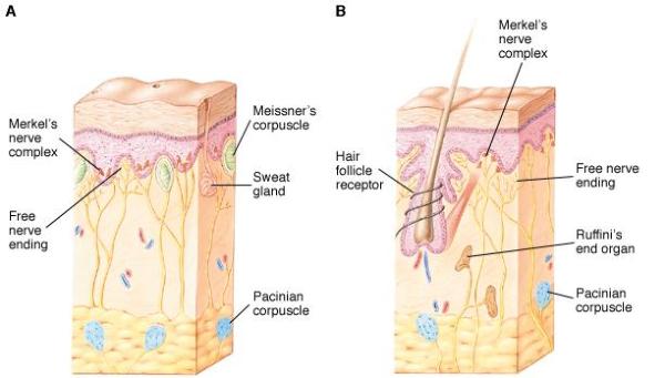
Peripheral Nerve Endings
Sensory Nerve Endings (Receptors)
The receptors are impulse detectors nerve endings or specialized cell.
They convert external or internal stimuli into afferent nerve impulse that pass to the CNS where they initiate appropriate response.
Classification of Sensory Nerve Endings
According to their locations
Exteroceptors
They are located at the body surface and detect external stimuli such as touch, light pressure, deep pressure, cutaneous pain, temperature, smell, taste, sight and hearing.
Proprioceptors
They are located within the skeletal system and provide information about orientation, skeletal position, tension and movement.
Such receptors include the vestibular apparatus of the ear, tendon organs and neuromuscular spindles.
Interoceptors
They are located within the visceral organs and respond to stimuli from viscera such as visceral pain, hunger, and thirst.
According to energy sensitivity
Mechanoreceptors
They are sensitive to mechanical distention such as distention of hollow organs (gastrointestinal tract and urinary bladder).
Chemoreceptors
They are sensitive to changes in the chemical composition of the blood.
Thermoreceptors
They are sensitive to changes in body temperature.
According to their morphology ![]()
![]()
Non-encapsulated sensory endings
Free Nerve Endings
They are non-myelinated small terminal branches of afferent nerve fibers found in the connective tissue and ramify among the epithelial cells throughout the body.
They detect sensory stimuli such as temperature, touch and pain.
Hair Follicle Terminals
They are touch receptors represented by free nerve endings derived from myelinated axon and extend among the epithelial cells of the hair follicles.
Non-encapsulated Tactile Corpuscle
They are touch receptors found along the dermo-epidermal junction of the skin.
The free nerve endings become associated with epithelioid tactile cells called Merkel cells scattered in the basal layers of the epidermis.
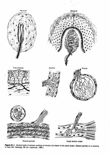
Encapsulated Sensory Nerve Endings
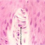
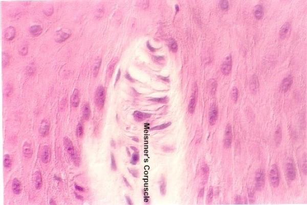
Encapsulated Tactile Corpuscle (Meissner’s Corpuscle) ![]()
They are oval-shaped touch receptors located in the dermal papillae.
They consist of delicate connective tissue capsule surrounding a mass of flattened, transversely arranged Schwann cells.
The non-myelinated terminal branches of one or two large myelinated axons ramify between the transverse cells.
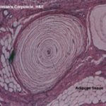
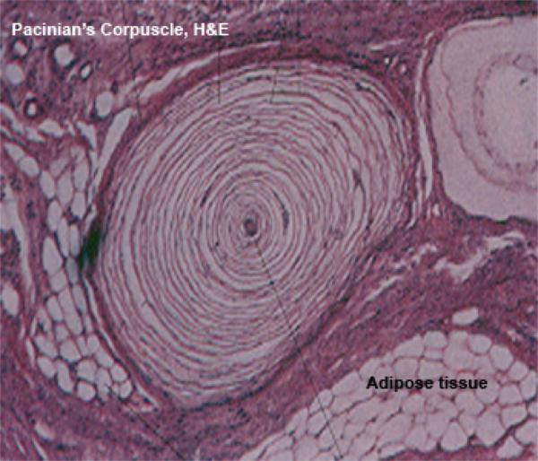
Lamellar, Pacinian or Vater Corpuscle ![]()
They are large encapsulated receptors (1-4 mm in length) found in the subcutaneous tissue, ligaments, joint capsule, pancreas, mesenteries and mammary glands.
They consist of delicate connective tissue capsule surrounding about 30 concentric lamellae of flattened cells separated by interstitial fluid and connective tissue fibers.
The center lamellae are densely packed around a single large non-myelinated axon.
They are sensitive to deep pressure or coarse touch.
End Bulb of Krause (Genital Corpuscle or Bulbous Corpuscle)
They are mechanoreceptors found in the conjunctiva, lips, dermis, glans penis and clitoris.
They consist of delicate connective tissue capsule surrounding highly coiled non-myelinated terminal branches that form a ball-like mass (glomerulus).
They are sensitive to touch and pressure sensation.
Golgi Tendon Organs (Neurotendineous Spindle)
They are found at the muscle-tendon junctions and are sensitive to tension of muscles.
They consist of thin connective tissue capsule surrounding unmyelinated axon that forms spray-like branches ending in small dilatations.
Ruffini Corpuscles
They are found in dermis, fascia, ligaments and joint capsule.
They are similar to neurotendenious spindle and are sensitive to mechanical distortion of the limbs.
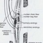
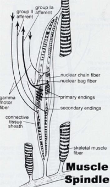
Neuromuscular Spindle (Muscle Spindle) ![]()
They are large (up to 6 mm long and less than I mm in diameter), encapsulated, lymph-filled, fusiform structures found within skeletal muscles.
Each spindle consists of 2-10 modified skeletal muscle fibers that are referred to as intrafusal fibers, in contrast to extrafusal fibers located outside the spindle.
There are two types of intrafusal fibers: nuclear bag fibers and nuclear chain fibers.
The nuclear bag fibers have dilated non-striated middle zone filled with nuclei.
The nuclear chain fibers are smaller than the nuclear bag ones and their nuclei are arranged in a single row.
The intrafusal fibers are associated with two types of sensory receptors: annulospiral endings and flower-spray endings.
The annulo-spiral endings are represented by non-myelinated sensory fibers of large myelinated sensory fibers that are wrapped around the nuclear region of the intrafusal muscle fibers.
The flower-spray endings are smaller myelinated sensory fibers and are located on the striated portion of the intrafusal fibers.
Information from muscle spindle is subconscious and is important for regulating muscle tone, adjusting posture and controlling movements.
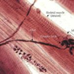
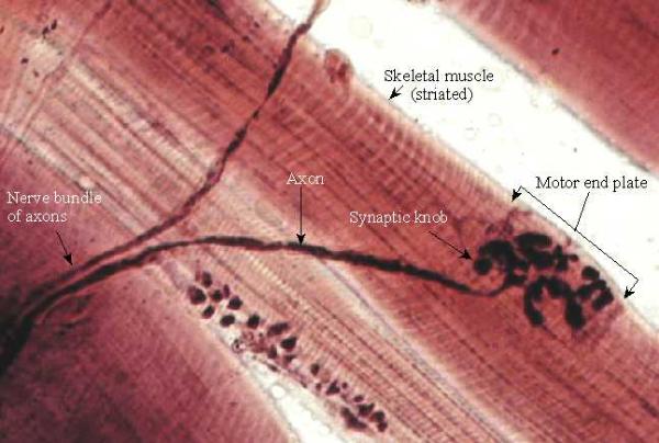
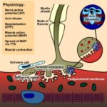
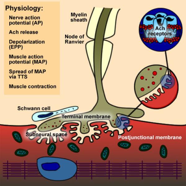
Motor nerve endings
Neuromuscular Junction ![]()
![]()
The neuromuscular junction is made up of motor-end plate, synaptic cleft, postsynaptic sole plate.
Motor End Plate
The terminal branches of the motor axon loose its myelin sheath and divides to form several small bulbous swellings called motor end plate.
Each end plate occupies a trough in the muscle cell surface (sole plate) and is covered by extension of the Schwann cell cytoplasm.
The end plate cytoplasm is rich in rER, mitochondria and clear synaptic vesicles containing acetylcholine.
Synaptic Cleft
It is a wide gap, about 30 mm separating the end-plate from the sole plate.
Sole Plate
It is the portion of sarcolemma that comes in contact with the end plate. It has many invaginations (troughs) that fit each end plate.
Each trough has several transverse enfolding called junctional folds to increase the surface area of sarcolemma.
Underneath the sarcolemma, the sarcoplasm is rich in mitochondria and aggregated nuclei.