-
- Figure 1

-
- Figure 2
-
- Figure 3
-
- Figure 4
-
- Figure 5
-
- Figure 6

-
- Figure 7
-
- Figure 8
-
- Figure 9
-
- Figure 10
-
- Figure 11
-
- Figure 12
-
- Figure 13
-
- Figure 14
-
- Figure 15

-
- Figure 16
-
- Figure 17

-
- Figure 18
Nervous tissue
The nervous tissue functions as a communication network to receive stimuli from both internal and external environment (exteroception) and subsequent transmission of signals or information (interoception) throughout the body or effector organs (muscles or glands). It is made up of nerve cells (neurons) and different types of supporting cells collectively known as neuroglia.
Neuron
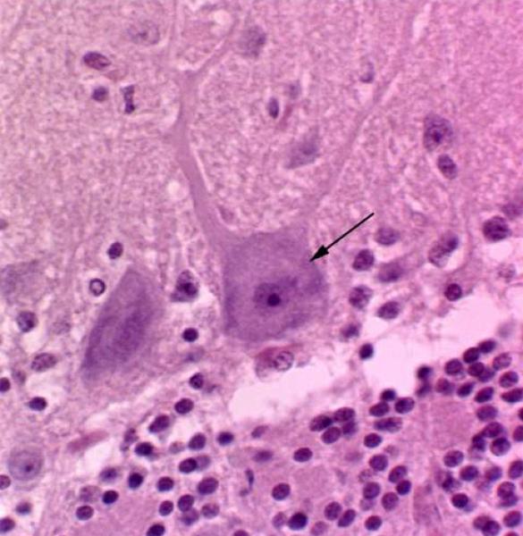
It is the structural and functional unit of the nervous tissue that is specialized in excitability and conductivity. Each neuron is composed of cell body (perikaryon) and cell processes. They are located in the gray matter of the central nervous system, eye (rods and cones), ears (organ of Corti), olfactory mucosa, and ganglia.
1) Cell body (perikaryon) ![]()
It includes the nucleus and it’s surrounding cytoplasm.
- Nucleus
The nucleus is large, spherical or ovoid and usually centrally located within the perikaryon, but in autonomic neurons it has an eccentric position. The chromatin is completely dispersed and the nucleolus is prominent (owl’s eye). With EM, a small amount of peripheral chromatin can be seen on the inner aspects of nuclear envelop. The female sex chromatin (Barr body) is usually evident either close to the nucleolus (cat) or the nuclear envelope (human being).
- Cytoplasm
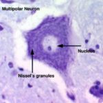
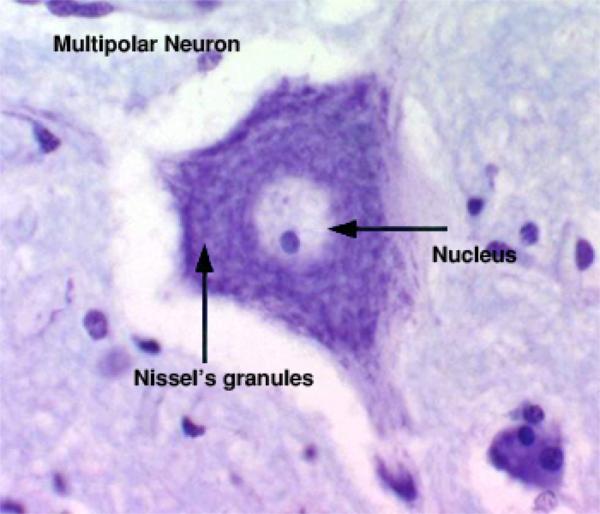
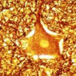
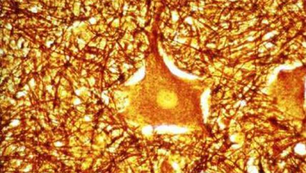
The cytoplasm of the nerve cells contains large aggregations of rER that appear as deeply basophilic masses scattered throughout the different cytoplasmic regions. These basophilic masses are called Nissl bodies or chromophilic substance ![]()
![]() and are especially prominent in large neurons. The great abundance of free ribosomes and polysomes that are located in the spaces between the rER cisternae is required to synthesis cytoplasmic protiens that continuously flow to replace proteins used up in metabolism.
and are especially prominent in large neurons. The great abundance of free ribosomes and polysomes that are located in the spaces between the rER cisternae is required to synthesis cytoplasmic protiens that continuously flow to replace proteins used up in metabolism.
In cases of neuronal injury, the perikaryon swells, the nucleus becomes eccentric and Nissl bodies disappear except peripherally (chromatolysis). This response is called axon reaction. The Golgi is diffuse and located mainly around the nucleus. Mitochondria are abundant, ovoid or thread like, and randomly distributed. Microfilaments and microtubules are numerous and arranged in parallel bundles throughout the perikaryon. Melanin pigments are found in the neuron of the substantia nigra in the midbrain. Lipofuscin are found especially in old neurons.
The neurons are highly differentiated cells. Centrioles are absent therefore they can not divide. Furthermore, from shortly after birth, new ones do not develop from precursor cells. The neurons synthesize neurotransmitter substances or their precursors in the perikaryon, they transported them along the axon to the synapse to be released when stimulated.
- Axon


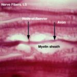
The axon is a long, cylindrical process of nearly uniform diameter and few branches. The axon originates from a region in the cell body called axon hillock that is a cone-shaped portion devoid of Nissl bodies.
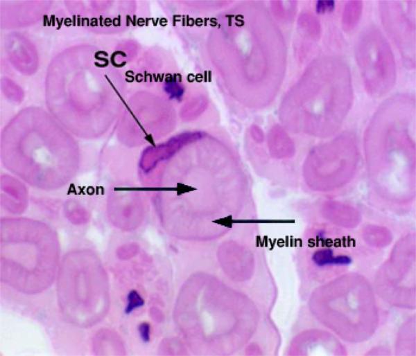
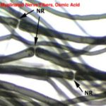
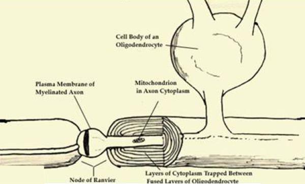
Some axons are invested by a segment of myelin sheath separated by gaps called nodes of Ranvier. The myelin sheath shows cone-shaped clefts (of Schmidt-Lantermann) that are actually helical tunnels extend from outside the sheath to inside. They are formed by portions of Schwann cell cytoplasm trapped within the myelin during its formation. Few collateral branches of the same diameter extend perpendicularly from the axon at nodes of Ranvier .
The region of the axon located just beneath the axon hillock is called initial segment. It is not invested with myelin sheath and at these region nerve impulses is initiated.
The axon is surrounded by plasmalemma (axolemma). The cytoplasm of the axon (axoplasm) is devoid of ribosomes and rER (Nissl bodies). It contains abundant neurofilaments and microtubules. In fixed state, a central band of parallel neurofilaments, which are continuous with those of the cell body, are found embedded in the axoplasm. Other organelles include numerous mitochondria and sER.
The axon ends in small branches called telodendria which end in small swelling called terminal buttons that contain synaptic vesicles and mitochondria and participate in synapse.
The axons are commonly refers as nerve fibers and they function as a major site for information output from the neuron.
- B) Dendrites
The dendrites are highly branched treelike, with broad origin and tapering ends with many fine spines called gemmules.
They are not myelinated and end either as specialized sensory receptors or form synapses with other neurons. They are the major sites of information input into the neurons. The main dendritic branch contains Nissl granules, mitochondria, microtubules and neurofilaments.
These organelles become progressively more scanty toward the ends of the dendrites. Synaptic vesicles are absent.
Mention the differences between axon and dendrites?
| Criteria | Axon | Dendrites |
| Contour | Regular contour and uniform diameter | Irregular contour with broad base and tapered ends |
| Number | Single | Multiple |
| Branching | Less branched, only few collateral branches | Highly branched |
| Nissl bodies | Absent | Present |
| Myelin sheath | Present | Absent |
| Function | Transmit nerve impulses away from the cell body | Transmit nerve impulses toward the cell body |
Classification of neurons
- According to the number of their processes
- Unipolar (Pseudounipolar) neurons

They are flask-shaped with a single large process that divides into two main branches, one of them acts as axon and the other acts as dendrites. (e.g., sensory neurons in craniospinal ganglia).
- Bipolar neurons

They are fusiform or spindle-shaped. Each cell has two processes, a single axon arises from one pole of the cell and a single dendrite arises from the opposite pole. (e.g., receptor neurons for the senses of smell, sight and balance).
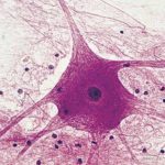
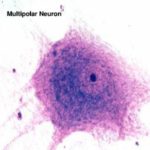
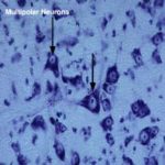
- Multipolar neurons


They are the most common forms. Each cell has a single axon and numerous dendrites. Dendrites may all arise from one pole of the cell body or may extend from all parts of the cell body. (e.g., neurons of CNS and autonomic ganglia).
- According to their shapes, three types of multipolar neurons are recognized:
- Polygonal (stellate neurons): are irregulars in shape with dendrites arise from different body regions (e.g., autonomic ganglia).
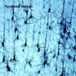
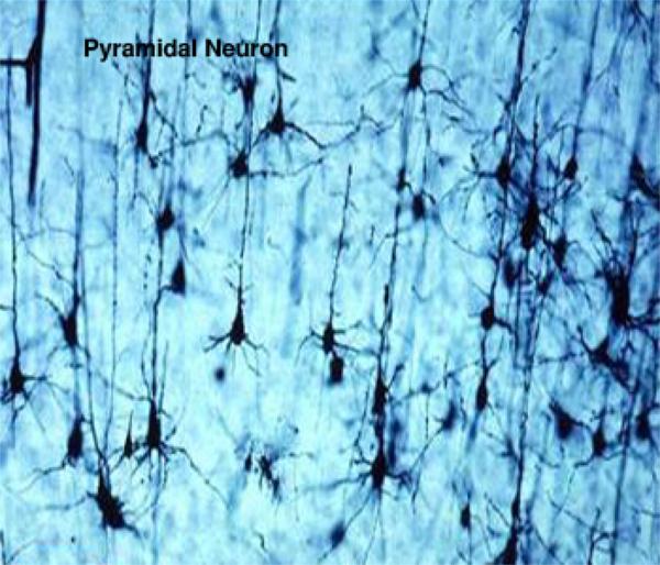
- Pyramidal neurons: are pyramidal in shape with dendrites arising from the apex and axon arises from its base (e.g., cerebral cortex).

- Pyriform neurons: are large, flask-shaped with extensively branched apical dendrites (e.g., Purkinje cells of the cerebellum).

- According to the length of their axons, two types are recognized:
- Golgi types I neurons: have long axons extend through peripheral nerves to reach their effector organs.
- Golgi types II neurons: have short axons usually end in the immediate vicinity of the cell.
Synapses
They are sites at which nerve impulses are either chemically or electrically transmitted from one neuron to another.
Classification of synapses
- According to the places at which the telodendritic process synapses on the postsynaptic neuron:
- Axodendritic synapse: occurs between axon and dendrite.
- Axosomatic synapse: occurs between axon and cell body.
- Axoaxonic synapse: occurs between tow axons.
- II) According to the presence of electron-dense granules on the cytoplasmic side of the postsynaptic membrane:
- Asymmetrical synapse: with postsynaptic density.
- Symmetrical synapse: has no postsynaptic density.
III) According to the way by which nerve impulses are transmitted:
- Chemical synapses: characterized by the presence of synaptic vesicles containing neurotransmitter substance within their telodendria. Nerve impulses induce the release of the neurotransmitter that diffuses across the intercellular space (synaptic cleft) to stimulate receptors in the postsynaptic membrane. Interaction of the neurotransmitter with postsynaptic receptors induces either excitation or inhibition of the other neurons.
- Electrical synapses: do not have synaptic vesicles and are similar to gap junctions with an intercellular cleft of about 2nm wide.
- Mixed synapses: use both electrical and chemical transmission.
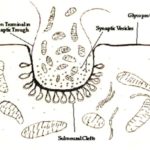
Structure of synapse ![]()
A synapse is made up of three main elements: presynaptic element, synaptic cleft and postsynaptic membrane.
- Presynaptic element
It consists of non-myelinated, dilated terminal portion of the telodendria.
It contains mitochondria and membrane-bound vesicles of neurotransmitter known as synaptic vesicles.
The synaptic vesicles are of two types; electron-dense (nor-epinephrine-containing) type and electron lucent (acetylcholine containing) type. They are derived from the sER of the axon.
The presynaptic plasmalemma has electron-dense plaques along its cytoplasmic surface. Synaptic vesicles tend to cluster around these regions. The presynaptic telodendria may assume one of the following shapes:
- Terminal bulbs or boutons: the axon terminates as bulbous enlargement.
- Passage synapse: some telodendria have multiple preterminal bulbs that form several synapses along its course.
- Calyx synapse: the telodendria are calyx-like and embrace the postsynaptic structure.
- Synaptic cleft
The presynaptic membrane is separated from the plasma membrane of the opposed neuron or effector cell by a narrow intercellular gap of uniform width (20-30 nm) called the synaptic cleft.
The cleft contains a protein-carbohydrate substance that may serve to hold pre-and-post-synaptic membranes in apposition.
- Postsynaptic membrane
It is the cell membrane of the adjacent neuron and it may have thicker accumulation of electron-dense substance. It is not associated with synaptic vesicles.
The cytoplasm beneath the postsynaptic membrane often contains a network of fine fibrils known as the postsynaptic web that forms desmosome-like structures that help to maintain the integrity of the synapse.
Neuroglia
They are a group of highly branched cells that occupy the spaces between neurons providing both mechanical and metabolic support. They are all derived from the ectoderm, except microglia that are mesodermal in origin.
Neuroglia of the CNS
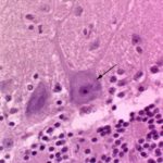
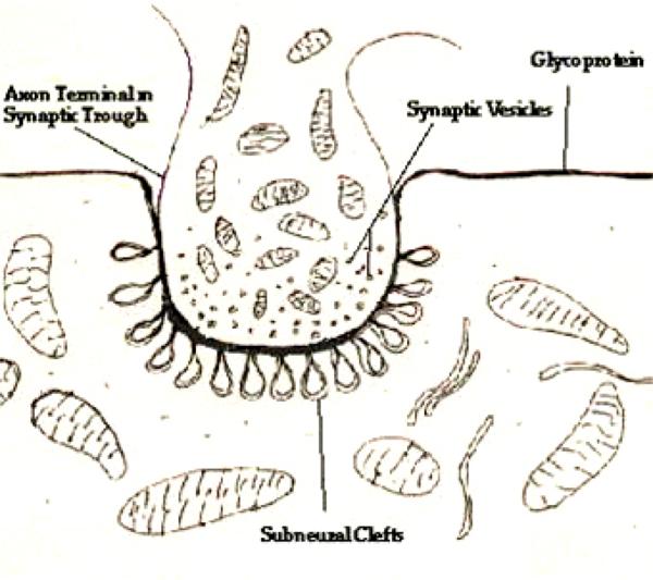
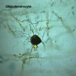
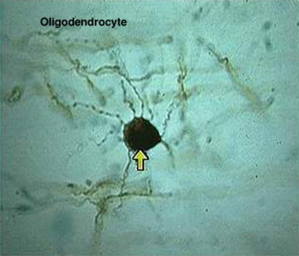
- Oligodendrocytes (Oligodendroglia) (Oligodendroglia, oligos, small, dendron, tree)
They are so named because they are fairly small with tree-like processes. ![]()
![]() In H&E sections, the cell has a small, spherical densely stained nucleus and a small number of thin, short-branched processes. The cytoplasm is rich in rER, mitochondria and microtubules.
In H&E sections, the cell has a small, spherical densely stained nucleus and a small number of thin, short-branched processes. The cytoplasm is rich in rER, mitochondria and microtubules.
Functions
- Formation of myelin sheathes around the CNS axons.
- A single cell may be responsible for the myelination of up to 50 nerve fibers.
3.They are usually found in rows between the myelinated fibers of the white matter.
- They surround neuron cell bodies in the gray matter.
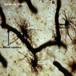
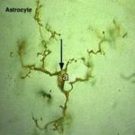
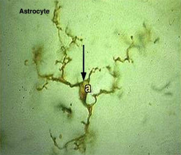
- Astrocytes (Astron, star)
They are so named because they possess radiating processes and have a star-like appearance. Two types are recognized:
- Protoplasmic astrocytes
They are the most numerous glial cells in gray matter. They have long, thick, highly branched processes that occupy most of the interneuronal spaces ![]()
![]() . The cytoplasm is rich in bundles of microtubules.
. The cytoplasm is rich in bundles of microtubules.
Many of the astrocyte processes terminate as end feet or perivascular feet upon the blood capillaries. Other processes terminate close to the non-synaptic regions of neurons.
- b) Fibrous astrocytes
They predominate in white matter. They have long, thin moderately branched processes. The cytoplasm is rich in microfilaments.
Functions
- Provide structural support for neurons.
2.Regulate the composition of the intercellular environment of the CNS. It takes up excess potassium when these escape from neurons during impulse conduction.
3.Mediate the exchanges of nutrients and metabolites between neurons and the blood.
- Forms diffusion barriers surrounding synapses.
- Phagocytosis.
- Scar tissue formation (gliosis) during the CNS injury.
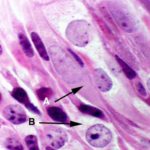
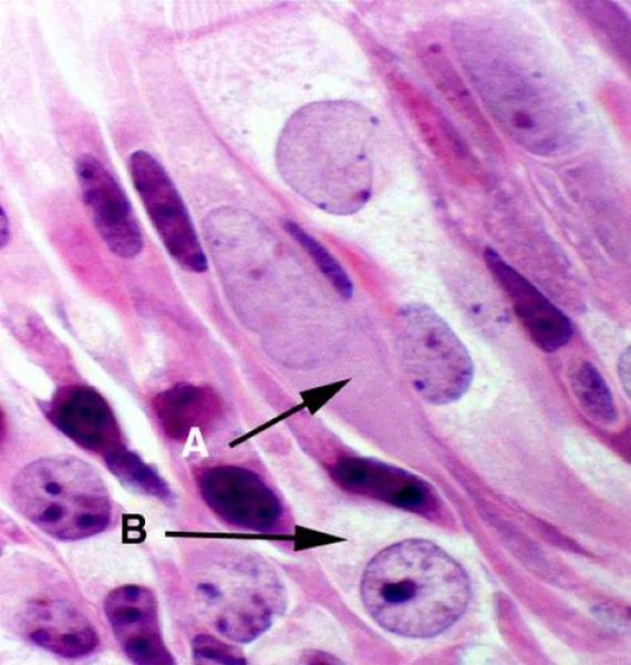
- Microglia
They are small cells evenly distributed in gray and white matter. They have little cytoplasm with fine highly branched processes. The nuclei are small, elongated and chromatophilic.
They are derived from the mesoderm. In normal tissue, the cells are sparse and difficult to found. In response to tissue damage, microglia transfer into large amoeboid phagocytic cells by which necrotic tissue is cleared away prior to gliosis.
In cases of CNS damage, several cell types may become phagocytic such as microglia, astrocytes, oligodendroglia, pericytes and blood macrophages.
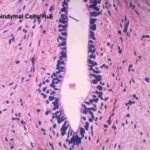
- Ependymal cells
Ependymal cells line the ventricles of the brain and the central canal of the spinal cord. They have a cuboidal or columnar shape and are tightly bound together at their luminal surfaces by zonulae adherents. Unlike the epithelia, these cells do not rest on a basement membrane. The cell bases from fine processes that interdigitate with the underlying layer formed by the processes of astrocytes. Their luminal surfaces have cilia and microvilli. ![]()
Function
- Production of cerebrospinal fluid.
Neuroglia of the PNS
- Neurolemmocytes (Schwann cells)
They are the supporting cells of the PNS. The primary function of these cells is the formation of myelin sheath around the myelinated axons. ![]()
In non-myelinated axons, each neurolemmocytes ensheathes a variable number of axons. Each axon is located inside an invagination within the surface of these cells.
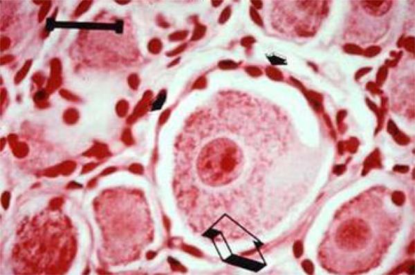
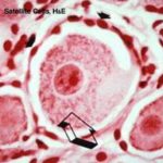
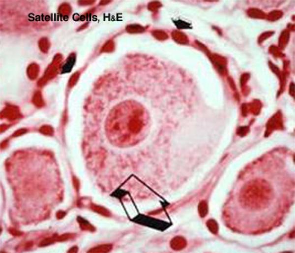
- Satellite cells
They are flattened neurolemmocytes that surround the neuronal cell bodies within the ganglia. ![]()