-
- Figure 1
-
- Figure 2
-
- Figure 3
-
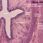
- Figure 4
-
- Figure 5
-
- Figure 6
-
- Figure 7
-
- Figure 8
-

- Figure 9
-
- Figure 10
-
- Figure 11
-
- Figure 12
-
- Figure 13
-
- Figure 14
-
- Figure 15
-
- Figure 16
-
- Figure 17
-
- Figure 18
-

- Figure 19
Female Reproductive System
The female reproductive system is formed of two ovaries, two uterine tubes, uterus, vagina and vulva as well as mammary glands.
The ovary is a combined exocrine (produce ova) and endocrine (produce estrogen and progesterone) gland.
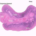
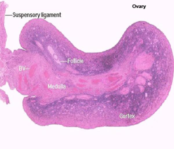
![]() The shape of the ovary varies among different animal species.
The shape of the ovary varies among different animal species.
It is oval or ovoid in cow, bean-shaped with a depression at one side (ovulation fossa) in mare and discoid highly trabercuated in sow.
The ovarian surface is covered by a single layer of cuboidal epithelium called the ovarian surface epithelium.
In mare, the ovarian surface epithelium is restricted to ovulation fossa, other areas are covered by mesothelium.
Beneath the surface epithelium, the outer limit of the ovary is demarcated by a tough, dense connective tissue, the tunica albuginea.
The tunica albuginea is made of collagen fibers, with a fair amount of smooth muscle.
The parenchyma of the ovary is divided into an outer cortex or zona parenchymatosa and an inner medulla or zona vasculosa.
In mare, the zona parenchymatosa occupies the central part and comes in contact with the periphery at the ovulation fossa.
The zona vasculosa occupies the periphery of the ovary except at the ovulation fossa.
The ovarian cortex contains the ovarian follicles and corpora lutea that are embedded in the cortical stroma.
The cortical stroma is essentially a ramification of the medulla, through which blood vessels enter and leave the cortex and surround the developing follicles.
The stromal cells have spindle nuclei and run in various directions surrounding the different cortical structures.
The medulla is formed of loose connective tissue contains nerves, many large coiled blood vessels, lymphatics, rete ovarii and smooth muscle.
In rodents, dog and cat ovaries, the cortical stroma contains interstitial gland cells.
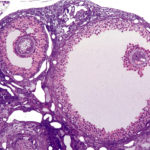
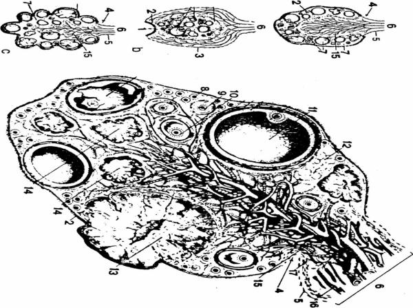
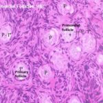
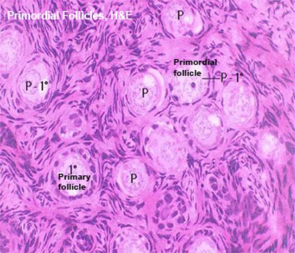
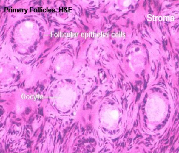
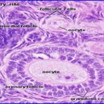
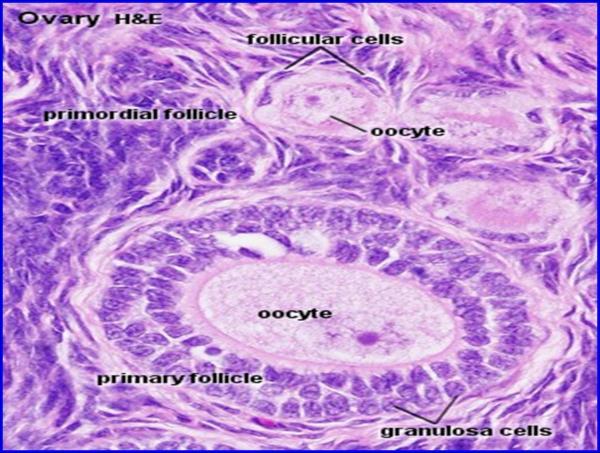
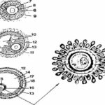
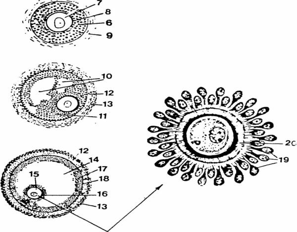
Ovarian Follicles ![]()
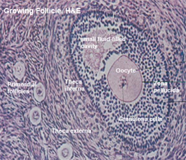
The primordial follicles, usually occurring in clusters.
This is the very earliest stage of development, and most follicles in any given ovary never get beyond this stage.
The primordial follicles consist of a large spherical primary oocyte that has a large lightly stained nucleus with prominent nucleolus surrounded by a single layer of squamous cells.
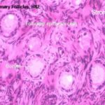
The primary follicles consist of a large primary oocyte surrounded by a single layer of cuboidal follicular cells.
N.B. The primary follicles are evenly distributed in ruminants and sow and occur in clusters in carnivores.
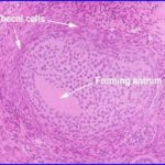
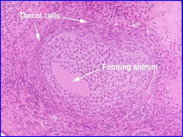
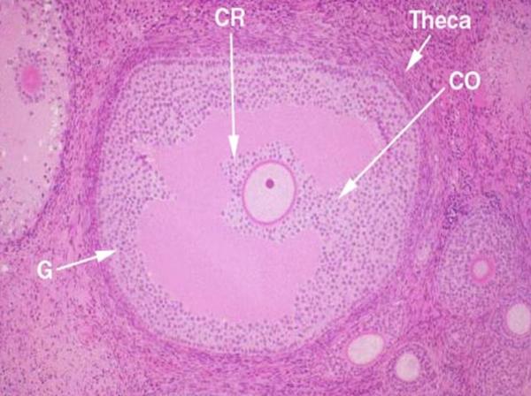
Secondary follicle ![]()
The secondary follicle consists of stratified follicular epithelium that surrounds primary oocyte and separated from it by zona pellucida.
The stroma begins to surround the secondary follicle forming theca folliculi.
The growing follicle has small fluid-filled cavities among the follicular cells. The theca folliculi is demarcated into theca interna and theca externa.
The mature follicle contains a large cavity or antrum, filled with follicular fluid, results from the coalescence of the small cavities.
The primary oocyte is displaced eccentrically in a thickened area of granulosa cells called the cumulus oophorus.
The granulosa cells are organized into zona granulosum that rests on a basement membrane. Some of the granulosa cells may contain large PAS – positive inclusions (Call-Exner bodies) which may represent precursors of liquor folliculi.
In the late stage of follicular development the follicular cells immediately surrounding the oocyte become columnar and radially disposed, they are then termed corona radiata.
The theca differentiates into two layers; theca interna and theca externa.
The theca interna are modified spindle shaped fibroblasts located in a delicate reticular connective tissue.
It contains an extensive blood and lymph capillary network.
In mature follicles, the cells become epithelioid and polyhedral. The cytoplasmic organelles in the epithelioid cells are typical of steroid-secreting cells. They secrete estrogens.
N.B. in rodent ovary, the epithelial theca interna cells of regressive follicle share in the formation of the ovarian interstitial gland cells.
The theca externa consists of loose connective tissue arranged around the theca interna.
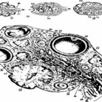
Ovulation ![]()
Ovulation is the process by which the oocyte, usually surrounded by the corona radiata is released from the mature follicle.
In the bitch and mare, both the first and the second meiotic divisions occur following ovulation, thus primary oocytes are liberated during ovulation.
In other animals, the first meiotic division occurs shortly before ovulation, thus secondary oocytes are liberated.
Atresia
Atrasia is the process by which follicles regress at any time during their development.
Atresia of primordial and primary follicles leaves no traces while atrasia of antral follicles leads to the formation of irregular scars, the atretic follicles.
In primary and secondary follicles, the oocyte degenerates before the follicular wall.
However, in mature follicles the reverse it trues.
Atretic mature follicles may include one of the following forms:
Obliterative atresia, in which the granulosa and theca layers may atrophy or only the granulosa layer may atrophy and the theca layer become either lutinize, fibrose or hyalinize.
Large cystic atretic follicle that do not regress, they interfere with the estrous cycle and fertility.
Signs of atresia include nuclear pyknosis, chromatolysis of oocyte nucleus.
The basement membrane may fold, thicken and hyalinize and is called glassy membrane.
Interstitial Gland Cells
They are found in rodent, bitch and queen.
They arise chiefly from the epithelioid theca interna cells of atretic antral follicles (follicles with antrum) or from hypertrophied granulosa cells of atretic preantral follicles.
The cells are polyhedral and contain large lipid inclusions.
Their ultrastructure features are suggestive for steroid-secreting cells.
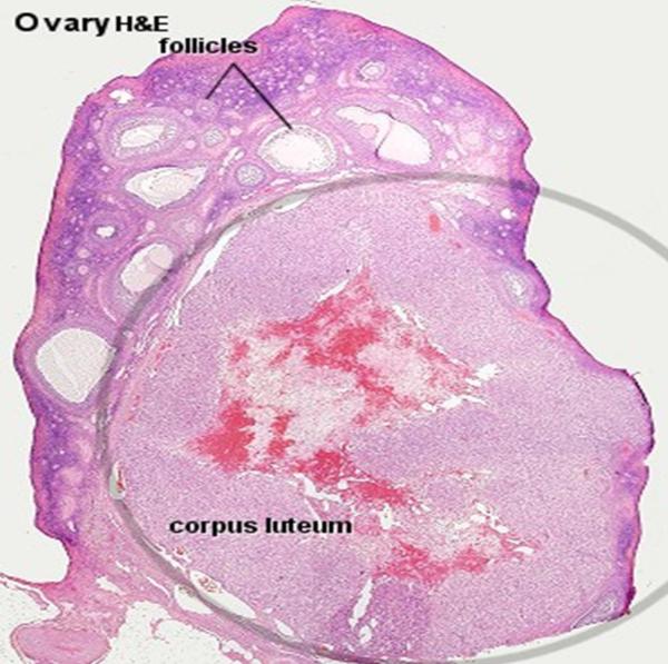
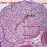
Following ovulation, the follicular wall of the ruptured follicle collapses.
The follicular cells are thrown into folds and protrude into the residual lumen that contains the blood that accompanies ovulation mixed with the remaining follicular fluid (corpus hemorrhagicum).
The granulosa cells enlarge, luteinize and are transformed into the large luteal (lutein) cell of the corpus luteum.
The fibroblast-type theca interna cells become spherical and contribute to the small luteal cell of the corpus luteum.
The corpus luteum is composed of cords and clumps of lutein cells supported by a delicate reticular network and demarcated into lobules by connective tissue septa.
The more central part still contains remnant of follicular fluid and clotted blood.
The corpus luteum is surrounded by connective capsule.
The lutein cells are differentiated into large and small lutein cells.
The large lutein cells are polygonal with spherical nuclei. Their cytoplasm contains cells organelles characteristic for steroid-secreting cells, lipid droplets and yellow lutein pigments in cow, mare and carnivores. They produce
progesterone.
The small lutein cells are found peripherally or as septa like clusters. They have more lipids and fewer steroid cells type organelles than large luteal cell.
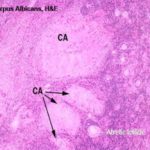
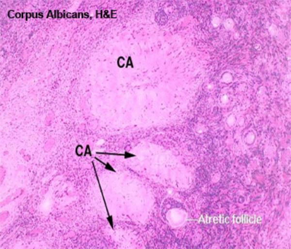
The connective tissue scar remaining after regression of ![]() corpus luteum is called corpus albicans.
corpus luteum is called corpus albicans.
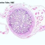
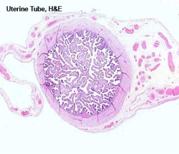
The oviduct is divided into three histologically distinct regions; infundibulum, ampulla and isthmus.
The wall of the uterine tube is formed of mucosa-submucosa, tunica muscularis and serosa.
The mucosa is highly folded with many primary longitudinal folds that are equipped with secondary and tertiary folds give the lumen of the ampulla the appearance of labyrinth of narrow clefts.
The epithelium is pseudostratified columnar with motile cilia on the majority of cells. Few non-ciliated secretory cells are also found among the ciliated cells.
The lamina propria-submucosa is a loose connective tissue layer with many plasma cells, mast cells and eosinophils.
The tunica muscularis consists chiefly of circular smooth muscle bundles but isolated longitudinal and oblique bundles also occur.
The serosa is a loose connective tissue with an outer mesothelium.
The mucosal folds are lower, less branched and less in number than ampulla. The epithelium is less ciliated and more secretory.
The muscular coat is thicker and consists of inner circular and outer longitudinal layers.
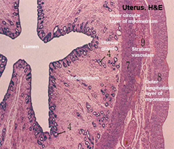
The uterine wall is formed of mucosa-submucosa (endometrium), tunica muscularis (myometrium) and serosa (perimetrium).
The epithelium is pseudostratified columnar and/or simple columnar.
The propria-submucosa is highly vascular, loose connective tissue with many fibroblasts, macrophages, mast cells, eosinophils, lymphocytes and plasma cells.
Simple branched tubular uterine glands whose distal ends have a variable degree of coiling are present.
The uterus of the ruminants characterized by the presence of dome-shaped prominences in the endometrium that are called uterine caruncles.
The caruncular areas are highly cellular, highly vascular and devoid of uterine glands. During pregnancy, caruncle (maternal part of the placenta) together with the cotyledon (fetal part of placenta) form placentome.
The uterine glands may be seen deeper to caruncular areas but their duct open in the intercaruncular regions
Uterine glands secrete uterine milk. In ewe, the caruncles are cup-shaped (i.e., dome with a central depression) containing many melanophores.
The tunica muscularis (myometrium) consists of a thick inner circular and an outer longitudinal layers of smooth muscle fiber, between the layers is a vascular layer consisting of large arteries, veins and lymph vessels called stratum
vasculare.
The perimetrium is a loose connective tissue covered by mesothelium.
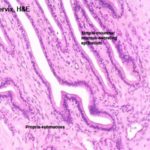
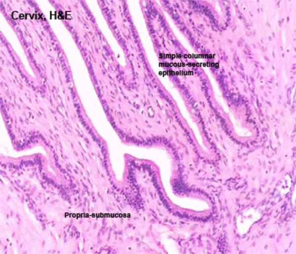
Cervix ![]()
The cervical mucosa is highly folded.
The epithelium is simple columnar with many mucigenous cells and goblet cells.
The lamina propria-submucosa consists of dense irregular connective tissue containing simple tubular glands in small ruminants and sow.
The tunica muscularis is formed of inner circular and outer longitudinal smooth muscle layers. Elastic fibers are prominent in the circular layer.
The serosa is a loose connective tissue layer with an outer mesothelial covering.
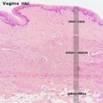
The epithelium is stratified squamous non-keratinized.
In cow a surface layer of columnar cells and goblet cells is present in the stratified squamous epithelium.
In bitch, intraepithelial glands are seen during estrus.
The lamina propria-submucosa is a loose or dense connective tissue rich in elastic fibers and lymphocytes.
The tunica muscularis consists of interlacing circular and longitudinal smooth muscle bundles. Longitudinally arranged bundles are more numerous.
The tunica adventitia is a loose connective tissue contains venous plexuses, nerve bundles and fat cells.
Vestibule and Valva
The wall of the vestibule is similar to that of the caudal portion of the vagina. In addition, more subepithelial lymphatic nodules are present.
The mucosa contains both major and minor vestibular glands.
The major vestibular glands are compound tubuloalveolar mucous glands in the deep part of the mucosa.
They occur in ruminants and queen. The glandular duct is lined by stratified squamous epithelium.
The gland has no capsule and is enclosed and partly divided by the striated musculature. They produce mucous for lubrication.
The minor vestibular glands are scattered in the vestibular mucosa of most domestic animals. In cow, they are concentrated in the median groove cranial to the clitoris.
Clitoris
The clitoris is located near the ventral commissar of the vulva.
It is rich in elastic tissue. It consists of corpus cavernosum clitoridis, glans clitoridis and preputium clitoridis.
The corpus cavernosum clitoridis is similar in its structure to the corpus cavernosum penis.
It is present in ruminants and well developed in the mare.
The glans clitoridis is the homologue of the glans penis and functionally erectile only mare.
The preputium clitoridis is a mucosal continuation.
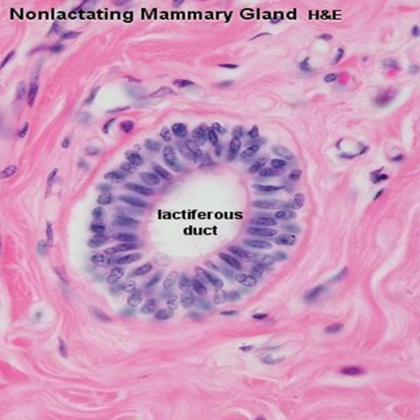
Mammary Gland
The mammary gland is a compound tubuloalveolar gland that is believed to be modified sweat gland.
It consists of udder and teats.
The udder consists of stroma and parenchyma.
The stroma of the udder is formed of dense fibroelastic capsule, fibrous septa and intralobular fine connective tissue.
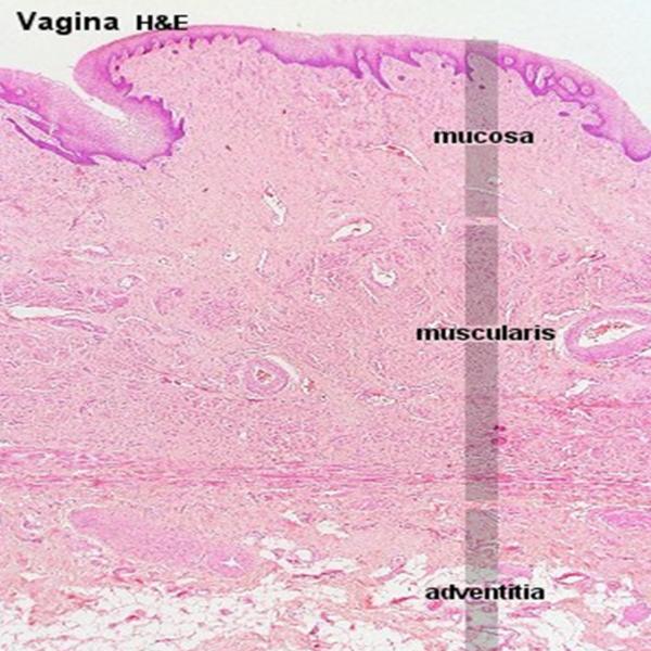
The parenchyma is formed of secretory mammary alveoli and excretory lactiferous ducts.
Actively lactating glands have much parenchyma and little stroma, whereas the reverse is true in nonlactating glands.
The lobules are packed with secretory alveoli and separated by thin connective tissue septa.
The secretory alveoli vary in appearance in different lobules of the mammary glands and are surrounded by reduced intralobular stroma.
In the synthesizing stage, the alveoli appear to be lined with high columnar cells with the free end protruding into the lumen.
In the exhaustion stage, the milk alveoli are lined with cuboidal epithelium.
- IRIS Readiris 11 Pro
- Macrabbit CSSEdit 2 MAC
- ARTS PDF Aerialist
- AutoCAD Structural Detailing 2015
- Pinnacle Studio 17 Ultimate
In-between the secretory alveoli there are intralobular ducts which are lined by simple cuboidal epithelium while in the septa there are interlobular ducts which are lined by stratified cuboidal epithelium.