-
- Figure 1
-
- Figure 2
-
- Figure 3
-
- Figure 4
-
- Figure 5
-
- Figure 6
-
- Figure 7
- II) Adult connective tissues
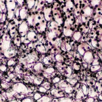
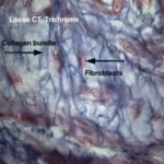
- Loose (ordinary or areolar) connective tissue

It is the most widely distributed type of connective tissue in the adult animals. It consists of all types of connective tissue cells, fibers that are embedded in non-sulfated amorphous ground substances. The cells are relatively more abundant than fibers that are loosely arranged leaving comparatively wide spaces in between.
The loose connective tissue is present around blood vessels and nerves and between muscle bundles. It supports the epithelial lining of gastrointestinal tract, respiratory and urinary tracts, also forms the deeper layers of skin and occurs as loose interstitial packing in many other organs.
- Dense connective tissue
The fibers are more abundant than cells and amorphous ground substances. According to the arrangement of its fibrous component, two types are identified:
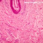
- Dense irregular connective tissue

It is formed of the same cell types like the loose connective tissue (all connective tissue cell type), although fibroblasts usually predominate, they are inactive with highly condensed nuclei and minimal cytoplasm.
The collagen fibers predominate, and they are arranged in coarse irregular interwoven bundles with very narrow space in-between.
It is found in lamina propria of the initial portion of the digestive tract, the capsule of the lung, the capsule of various organs (spleen, liver, kidney, testis), fascia, joint capsule and dermis.
- Dense regular connective tissue
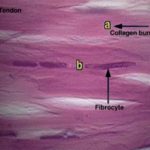
- Dense regular collagenous (tendon)

The tendon consists of bundles of parallel collagen fibers that are bounded together by sparse, loose connective tissue contains small blood vessels, nerves and active fibroblasts (peritenteneum interna).
The peritenteneum interna is continuos with the peritendineum externa that is a loose connective tissue capsule that cover the outer surface of the tendon.
The active fibroblasts that are located in the loose connective tissue layer between the bundles are responsible for the repair of tendons whenever the needs arise.
The fibrocytes located between the collagen fibers are inactive cells and appear as long, flat cells with wing-like cytoplasmic processes extending between adjacent collagen fibers, giving them a stellate appearance (bird cells) in cross sections.
- Dense regular elastic (Elastic ligaments)
It consists of branching and interconnected parallel elastic fibers surrounded by loose connective tissue (e.g., ligamentum nuchae and the elastic fascia of the abdominal muscle of herbivores).
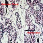
- Reticular connective tissue

It is made up of stellate reticular cells and a complex network of delicate thin branched and anastomosed reticular fibers.
It forms a delicate supportive framework for many highly cellular organs such as endocrine glands, liver and lymphoreticular organs (tonsils, spleen, and lymph nodes).
- Adipose tissue
It is a special type of connective tissue designed to perform
many functions such as mechanical protection, thermal
insulation and body metabolism. There are two types of adipose
tissue white and brown adipose tissues.
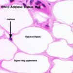
- White adipose tissue

It is distributed throughout the body especially in the deep layer of the skin and around the kidneys. White fat is divided by septa of loose connective tissue into clusters of adipose cells known as lobules. A delicate network of collagen and reticular fibers that support a dense capillary plexus and nerve fibers surrounds each adipocyte. In addition, the narrow intercellular spaces contain a few fibrocytes, mast cells, and scanty amorphous ground substance.
Fat stored in adipocytes as single large droplet (monolocular fat cell) which occupies most of the cytoplasm. The nucleus is compressed and displaced to one side of the cytoplasm giving the cells their characteristic signet ring appearance.
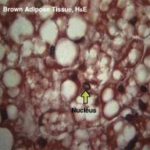
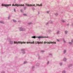
- Brown adipose tissue

It is a highly specialized form of adipose tissue found in newborn mammals, rodents and some hibernating animals, where it plays an important part in body temperature regulation.
The brown adipose tissue is more vascular than the white ones. The brown adipocytes are smaller than white one, the nuclei are oval, eccentricity located and surrounded by a significant amount of strongly acidophilic cytoplasm.