-
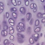
- Figure 1
-

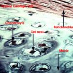
- Figure 2
-
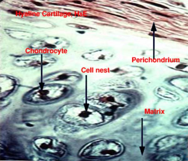
- Figure 3
-
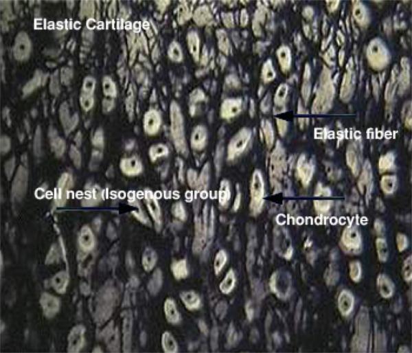
- Figure 4
Cartilage
The cartilage is a semi-rigid, flexible, avascular form of connective tissue designed to perform a supportive function. It is composed of cells and matrix (fibers and amorphous ground substance).
Types of cartilage
- Hyaline cartilage

The hyaline cartilage consists of:
- Perichondrium
It is a vascular connective tissue capsule that invests the external surface of cartilage. It is composed of two layers: a) Outer fibrous layer, composed of dense irregular connective tissue containing blood vessels and nerves. b) Inner cellular or chondrogenic layer made up of chondroblasts that are actively involved in production of matrix during cartilage growth and regeneration.
- Cartilage cells
- Chondroblasts (cartilage forming cells)
They are found mainly in the inner chondrogenic layer of the perichondrium. They are oval or spindle-shaped cells with oval euchromatic nuclei. The cytoplasm is basophilic rich in ribosomes, rER and Golgi saccules. They secrete matrix around themselves and become deeply buried in the cartilage matrix where they are called chondrocytes.
- Chondrocytes (mature cartilage cells)
They are located in tiny spaces within the cartilage matrix known as lacunae.
Beneath the perichondrium, chondrocytes are small and their lacunae are elliptical with their long axes parallel to the surface.
Deep within the cartilage, the cells are larger and polyhedral with short processes. They have a spherical nucleus wit one or mare nucleoli. The cells accumulate glycogen and lipid in their cytoplasm those appear vacuolated.
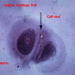
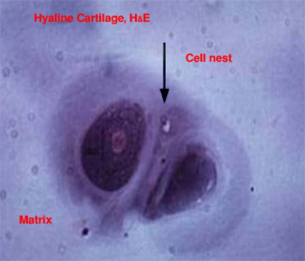
Some lacunae contain only one cell; others contain two, four, or sometimes six cells. These multicellular lacunae are called cell nests or isogenous groups because each cluster is the progeny of one cell.
- Matrix
The hyaline cartilage matrix is an amorphous gel consists mainly of sulfated glycosaminoglycans that are strongly basophilic, PAS positive and metachromatic.
The fibrous component represented by fine collage fibrils made up of type II collagen that has the same index of refraction as the amorphous ground substance, therefore, they can not be seen in common H&E sections.
The hyaline cartilage occurs in many places such as articular surface, fetal skeleton, nasal septum, larynx, trachea and bronchi.
- Elastic cartilage

The histological structure of the elastic cartilage is similar to that of the hyaline cartilage except: 1) cell nests are few. 2) The matrices contain a dense network of elastic fibers that are visible in H&E sections.
The elastic cartilage occurs in the external ear and external auditory canal, the epiglottis, corniculate and cuneiform cartilage of the larynx.
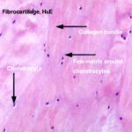
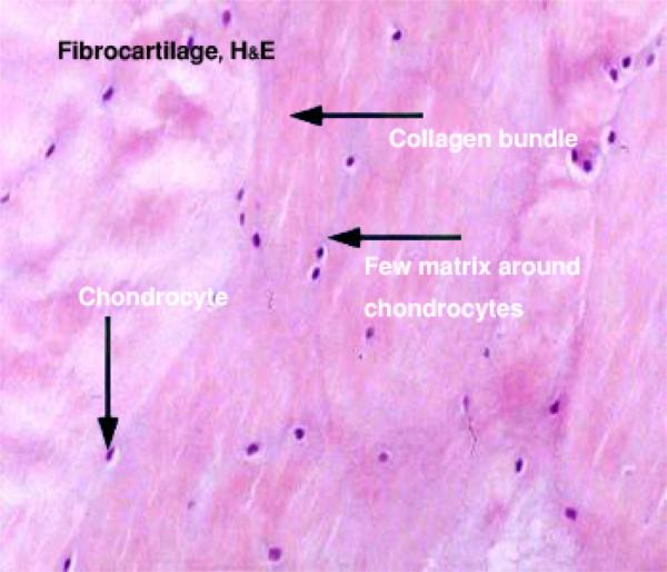
III. Fibrocarlilage ![]()
It is a transitional form between hyaline cartilage and dense regular connective tissue. It consists of regular parallel bundles of collagen fibers separated by encapsulated chondrocytes that occur singly, in pairs or sometimes form rows. The ground substances are little and only found around the chondrocytes. The fibrocartilage is found in the intervertebral discs, menisci of the stifle joint and at the attachment of tendons and bones.
Cartilage growth
- Appositional growth
In this type of growth, the chondroblasts of the inner chondrogenic layer of the perichondrium divide and gives rise to new daughter cells.
The cells begin to secrete matrix around them forming new cartilage layer that is added to the periphery of the cartilage. These cause the cartilage to grow in width.
- Interstitial growth
The chondrocytes located inside their lacunae start to divide forming cell nests (isogenous group) ![]() . The matrix produced by each daughter cells cause the cartilage matrix as a whole to expand from within. This causes the cartilage to grow in length.
. The matrix produced by each daughter cells cause the cartilage matrix as a whole to expand from within. This causes the cartilage to grow in length.
Cartilage nutrition
The cartilage is avascular connective tissue type. The cartilage cells receive their nutritional support mainly by diffusion of oxygen and nutrients through the matrix from blood vessels located within the perichondrium.
Calcification of the cartilage matrix may occur in aging and during bone development. In this case, diffusion is blocked and the cartilage cells die.