-
- Figure 1
-
- Figure 2
-
- Figure 3
-
- Figure 4
-
- Figure 5
Glandular epithelium
Classification
- According to the presence or absence of ducts
- Exocrine glands
They are glands that have a duct system to convey their secretory products to the sites of utilization (e.g., salivary glands).
- Endocrine glands
They do not have a system of duct (ductless). The secretory product (hormone) reaches the site of utilization through blood or
lymph (e.g., pituitary gland and thyroid gland).
2) According to the number of cells forming the gland
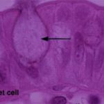
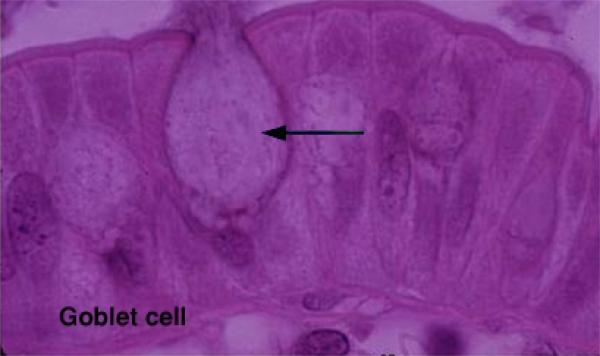
- Unicellular glands

It consists of a single secretory cell in a non-secretory epithelium (e.g., goblet cells).
- Multicellular glands
It is composed of more than one cell (e.g., salivary gland).
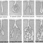
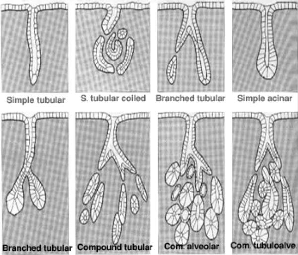
3) According to the morphology of duct and the secretory parts ![]()
- Simple tubular where the duct is not branched and the secretory part is in the form of tubule (e.g., glands of the large intestine).
- Simple acinar or alveolar glands where the duct is not branched and the secretory part is in the form of alveolus or acinus (e.g., sebaceous gland and the glands of skin of amphibians).
- Simple tubuloalveolar glands where the duct system is not branched and the secretory part is tubular and
alveolar (are rare).
- Simple branched tubular where the duct is not branched while the tubular secretory part is branched (e.g., glands of the stomach).
- Simple branched alveolar where the duct is not branched while the alveolar secretory part is branched (e.g., sebaceous glands).
6.Simple branched tubuloalveolar where the duct is not branched while the tubular and alveolar secretory part is branched (e.g., minor salivary glands).
7.Compound tubular glands where the duct is branched and the secretory parts are tubular (e.g., liver).
8.Compound alveolar glands where the duct is branched and the secretory parts are alveolar (e.g., mammary
glands).
9.Compound tubuloalveolar glands where the duct is branched and the secretory parts are tubular and alveolar
(e.g., salivary glands and pancreas).
5) According to the nature of secretion
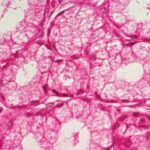
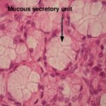
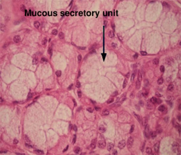
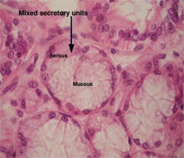
They produce thick, viscous secretions (mucus). The cells of the mucous secretory units are cuboidal in shape and
filled with mucinogen, the precursor of mucus that stain light (foamy or vacuolated) in H&E.
The nuclei are flattened and rest on the basement membrane. The lumen is wide (e.g., palatine glands and the glands of the tongue).
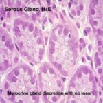
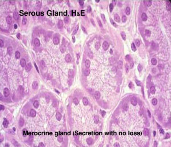
They produce thin watery secretion.
The cells of the secretory units are pyramidal in shape. The nuclei are spherical and situated near the center of the cells.
The cytoplasm has two zones, basal zone that appears basophilic due to the presence of rER and apical eosinophilic zone due to the presence of zymogen granules. (e.g., parotid glands and pancreas).
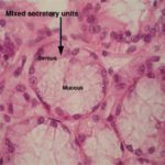
- Seromucoid or mixed glands

They produce mixed secretions.
They consist primarily of mucous secretory units with crescent-shaped clusters of serous cells (serous demilunes)
located at the periphery of the mucous units.
The serous secretion reaches the lumen through intracellular canaliculi located between the mucous cells. (e.g., submandibular and sublingual salivary glands).
6) According to the mode of secretion
- Merocrine glands (secretion without loss)

The cells of which remain intact and not destroyed during the process of secretion. The secretory granules are
discharged by exocytosis (e.g., salivary glands).
The apical parts of the cells are destroyed during the secretory process (e.g., some sweat glands and
mammary glands).
The whole secretory cells are discharged then destroyed to release the secretory product (e.g., sebaceous glands).