-
- Figure 1
-
- Figure 2
-
- Figure 3
-
- Figure 4
-
- Figure 5
-
- Figure 6
-
- Figure 7
-
- Figure 8
-
- Figure 9
-
- Figure 10
-
- Figure 11
Membrane specialization of epithelia
The basal, luminal and intercellular surfaces of epithelial cells have a variety of specializations.
- Basal surface
- Basement membrane
The epithelium is separated from the underlying connective tissue by a thin membrane known as basement membrane ![]() . It consists of two layers; basal lamina and reticular lamina.
. It consists of two layers; basal lamina and reticular lamina.
The basal lamina is synthesized by the adjacent epithelial cells and is located in contact with the epithelial basal plasmalemma. It is composed of type IV collagen embedded in an amorphous matrix of structural glycoprotein called laminin.
The reticular lamina is derived mainly from the underlying connective tissue and is located deep to the basal lamina. It consists of fine reticular fibers embedded in an amorphous ground substance.
In addition to underlying all epithelia, a basal lamina is found around muscle cells, neurolemmocytes and between epithelia in the renal corpuscle.
The basement membranes are difficult to resolve in common H&E sections, however, they can be selectively stained with silver (black) or PAS (magenta color).
Functions
- Support epithelial surfaces.
- It may act as selective filter, such as the glomerular basement membrane of the kidneys.
- It acts as a selective barrier to passage of cells between epithelia and connective tissue. For example, they permit the passage of the immune cells but prevent epithelial and connective tissue cells.
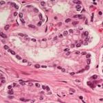
- Hemidesmosome
It is a half-desmosomes that is found at the interface between stratified squamous epithelium and the basement membrane.
- Basal surface enfolding
- I Apical surface
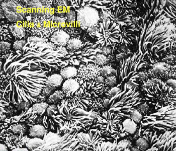
The luminal surface may have cilia, microvilli or stereocilia.
- Cilia
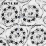
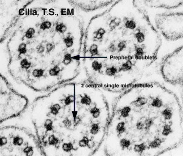
They are motile structures about 7-10 mm in length, which project from certain epithelial surface such as those of the respiratory and female reproductive tracts. They are much longer than microvilli and are readily visible with LM.
![]() With EM, each cilium is bounded by an evagination of the luminal plasma membrane and contains a central core called the axoneme. An axoneme consists of two central single microtubules surrounded by nine peripheral doublets. The microtubules of a doublet (subunit A and B) share a common wall of two to three protofilaments. Adjacent doublets are linked to each other via protein bridges called nexins and are also linked to the central sheath by radial spokes. On each doublet, there are short dynein arms projecting from subunit A
With EM, each cilium is bounded by an evagination of the luminal plasma membrane and contains a central core called the axoneme. An axoneme consists of two central single microtubules surrounded by nine peripheral doublets. The microtubules of a doublet (subunit A and B) share a common wall of two to three protofilaments. Adjacent doublets are linked to each other via protein bridges called nexins and are also linked to the central sheath by radial spokes. On each doublet, there are short dynein arms projecting from subunit A ![]() .
.
At its base, the axoneme inserted into a structure called a basal body that is similar in its structure to that of a centriole,
Functions
- In the respiratory airways, cilia beat in wave-like rhythm propelling mucus secreted by goblet cells towards the throat where it is swallowed thus keeping the airways clean.
- In the oviducts, ciliary action plays a part in transporting the ovum from the ovary towards the uterus.
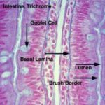
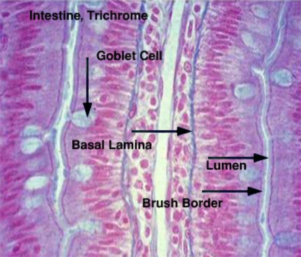
- Microvilli
![]() They are minute finger-like projections of the luminal plasma membrane found in many epithelial cells particularly those specialized for absorptive function (i.e. intestinal epithelium). Individual microvilli are too small (0.5-1 mm in length) to be resolved by LM. In certain cells such as in the small intestine and proximal renal tubules, the microvilli are so numerous and regularly arranged that under the LM the cell surface appears striated and is called striated or brushes border.
They are minute finger-like projections of the luminal plasma membrane found in many epithelial cells particularly those specialized for absorptive function (i.e. intestinal epithelium). Individual microvilli are too small (0.5-1 mm in length) to be resolved by LM. In certain cells such as in the small intestine and proximal renal tubules, the microvilli are so numerous and regularly arranged that under the LM the cell surface appears striated and is called striated or brushes border.
With EM, the core of each microvillus contains actin filaments that insert into the terminal web that is specialization of the cytoskeleton lying immediately beneath the cell surface. At the tip of each microvillus, the filaments attach to an electron-dense part of the plasma membrane.
Function: increase the surface area for absorption.
- Steriocilia
They are extremely long, branched microvilli. They are non-motile and readily visible with LM on the epithelium lining the epididymis. It facilitates absorption through increase the surface area.
III. Intercellular surfaces (lateral surfaces)
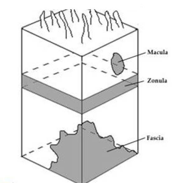
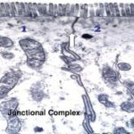
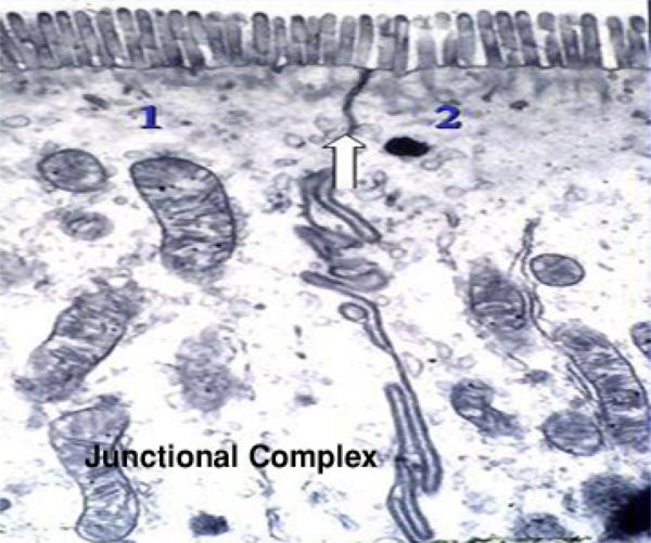
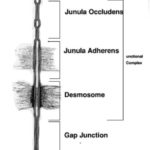
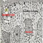
Cell junctions and junctional complex
![]() The cell junction holds epithelial cells together and is composed of four different types:
The cell junction holds epithelial cells together and is composed of four different types:
- Zonula occludens (tight junctions)


It is located just below the luminal surface and consists of small areas where the outer laminae of opposing plasma membranes are fused with each other.
As the term zonula (belt-like) implies, the tight junction forms a complete circumferential belt around each cell thus sealing the intercellular space from the lumen.
- Zonula adherens
It is located deep to the tight junctions forming a circumferential belt (zonula) around each cell. At these areas, the opposing plasma membranes diverge; no structures are evident between the opposing cell membranes. On the cytoplasmic aspect of these junctions, there is a fine network of filamentous materials that fuse with the filaments of the terminal webs.
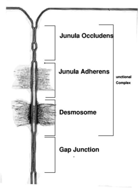
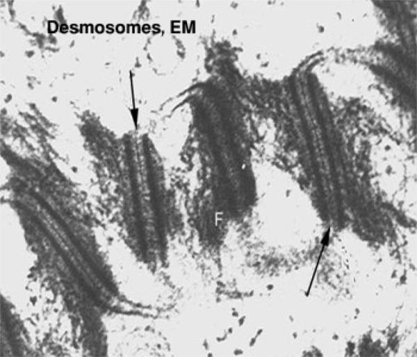
- Macula adherens (desmosomes)

The term macula indicates that the junctional area is in the form of spot-like.
It constitutes the third component of the junctional complex but also occurs singly at many intercellular sites.
At the desmosome, the opposing plasma membranes are separated by a gap in which several fine, transverse filaments or dense longitudinal line may be seen. At the cytoplasmic aspect of each plasma membrane, there is an electron-dense layer into which fibriller elements of the cytoskeleton appear to converge.
- Gap junctions
At these areas the opposing plasma membranes are very close to one another, but there is no fusion occurs. They are still separated by a narrow gap.
The Gap junctions function primarily as a passageway for ions and small molecules between adjacent cells. Thus, it is responsible for metabolic coupling and functional synchronization between adjacent cells (smooth and cardiac muscle).
Alteration of cell constituents in correlation to function
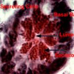
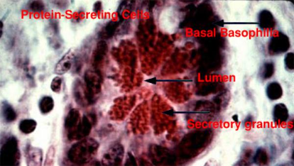
- Protein secreting cells
![]() The protein secreting cells are characterized by the presence of rough endoplasmic reticulum with abundant fixed ribosomes and polyribosomes, many mitochondria, well-developed Golgi stacks and less electron dense nucleus with extended chromatin. Zymogen granules are being discharged at the secretory surface of the cell. The unit membrane of secretory vesicles being added to cell membrane.
The protein secreting cells are characterized by the presence of rough endoplasmic reticulum with abundant fixed ribosomes and polyribosomes, many mitochondria, well-developed Golgi stacks and less electron dense nucleus with extended chromatin. Zymogen granules are being discharged at the secretory surface of the cell. The unit membrane of secretory vesicles being added to cell membrane.
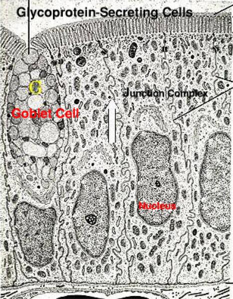
- Glycoprotein-secreting cells
The glycoprotein secreting cells have abundant basal rER, well-developed Golgi and numerous secretory granules that are extruded at the free surface of the cell by exocytosis.
The ion transporting cells have multiple and deep invaginations of the basal lamina being associated with abundant vertically oriented mitochondria.
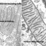
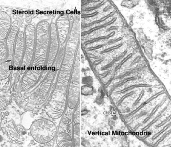
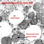
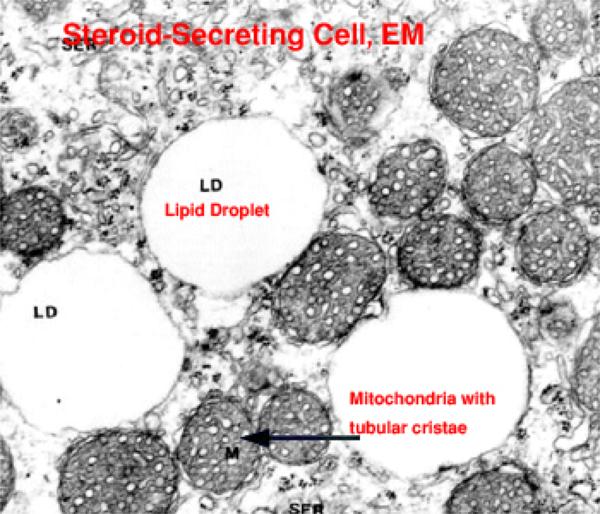
- Steroid secreting cells
![]() The steroid secreting cells have abundant sER, numerous mitochondria with tubular cristae and many lipid droplets.
The steroid secreting cells have abundant sER, numerous mitochondria with tubular cristae and many lipid droplets.