-
- Figure 1
-
- Figure 2
-
- Figure 3
-
- Figure 4
- Stratified epithelium
It consists of tow or more than tow layers of cells.
- Stratified squamous epithelium
It consists of several layers of cells with only the superficial cells having squamous shape.
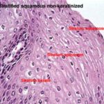
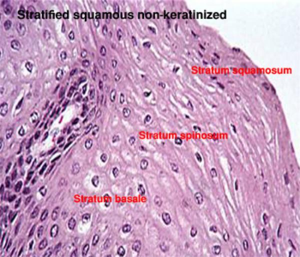
- Stratified squamous non-keratinized epithelium

It consists of three layers:
1.Stratum basale is a single layer of cuboidal to columnar cell rest on a wavy basement membrane.
- Stratum spinosum is composed of several layers of polyhedral cells tightly adhere to each other by numerous desmosomes. In H&E sections, the desmosomal attachments appear as small spiny processes, hence the name of this layer (spiny layer or stratum spinosum). The stratum basale and the deep layer of stratum spinosum are involved in active mitosis, therefor this region is referred to as stratum germinativum.
- Stratum squamosum is the superficial layer and is made up of few layers of flat squamous cells with ovoid small nuclei.
Locations: oral cavity, pharynx, esophagus, anal canal, and vagina. Such sites are normally subjected to moderate mechanical abrasion and are kept moist by local glandular secretions.
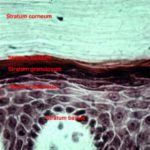
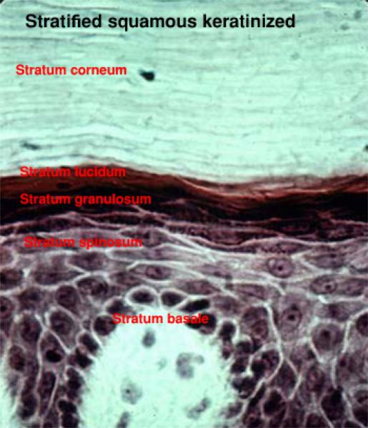
It consists of five layers:
- Stratum basale: consists of single layer of cuboidal to columnar cells resting on a wavy basement membrane.
- Stratum spinosum: has the same structure like that of the stratified squamous non-keratinized epithelium.
- Stratum granulosum consists of few layers of flattened cells having small pyknotic nuclei and rich in keratohyaline granules.
- Stratum lucidum found only in non-hairy skin. It is a layer of flattened, keratinized cells between the stratum granulosum and stratum corneum. It has a translucent or shiny appearance because the cytoplasm of these cells is rich in protenecious materials called eleidin.
- Stratum corneum is the outermost layer and consists of dead, keratinized cells. The cells have lost their nuclei and their cytoplasms filled with keratin that is a water-resistant protein.
- Stratum disjunctum formed of groups of cells in the outermost layer of the stratum corneum that become loose and detach to constitute this layer.
Locations: epidermis, hoof and horns.
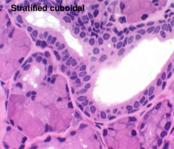
- Stratified cuboidal epithelium

It consists of two or more layers of cells, with only the superficial cells having a cuboidal shape. It is frequently occurs as two-layered epithelium located in the large glandular ducts.
It consists of several layers of cells with only the superficial layer having tall columnar cells.
Locations: distal portion of the urethra, parotid and mandibular ducts, lacrimal sac and lacrimal duct.
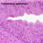
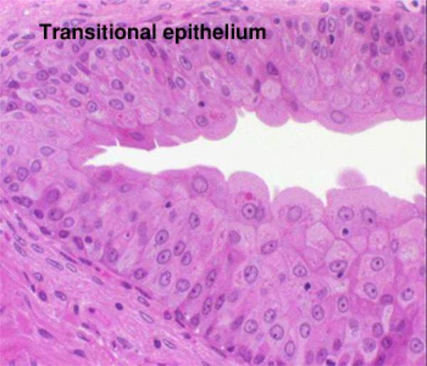
It is a form of stratified epithelium found only in the urinary tract (lines the ureter and urinary bladder). It is highly specialized to resist a great degree of stretch and to withstand the toxicity of urine.
In relaxed state (empty bladder) it is consists of 4-5 layers of cells, the basal layer is cuboidal in shape rests on thin basal lamina . The intermediate layer consists of several layers of polyhedral or pear-shaped cells. The surface cells are large cuboidal or dome-shaped with convex outer surface and concave inner surface. Their nuclei are large, spherical with prominent nucleoli, some cells are binucleated.
In stretched state (full bladder) it appears only as two or three cell layers thickness. The intermediate and surface layers are extremely flattened. The superficial cells have a thicker plasmalemma that acts as a barrier against diffusion of fluid from the subepithelial tissue to the hypertonic urine.